
Fig 5
- ID
- ZDB-IMAGE-220307-5
- Publication
- Habicher et al., 2022 - Chondroitin/dermatan sulfate glycosyltransferase genes are essential for craniofacial development
- All Figures
- Figures for Habicher et al., 2022
|
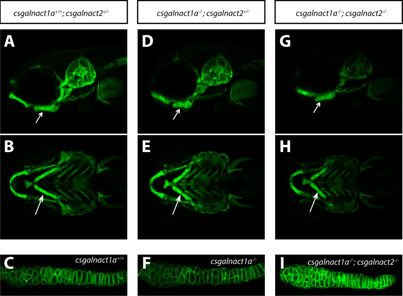
Fig 5
LightSheet images of live Tg(col2a1a:mEGFP) larvae at 6 dpf show craniofacial cartilage elements formed in csgalnact1a+/+;csgalnact2+/- (A,B,C), csgalnact1a-/-;csgalnact2+/- (D,E,F) and csgalnact1a-/-;csgalnact2-/- larvae (G,H,I). Lateral views (A,D,G) display a shorter ceratohyal, ventral views display a wider angle between the ceratohyal elements in csgalnact1a-/-;csgalnact2+/- (E) and csgalnact1a-/-;csgalnact2-/- (G) larvae (arrow) compared to controls (A,B). A detailed view on the chondorcytes within the ceratohyal (arrow) shows that intercalation occurs even in csgalnact1a-/- (F) and csgalnact1a-/-;csgalnact2-/- (I) larvae.