
Image
|
Figure Caption
FIGURE 2
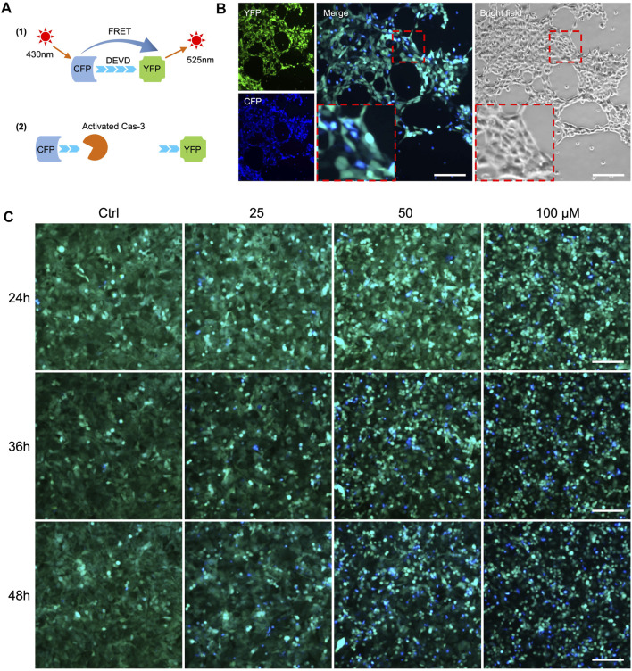
FRET-based detection of drug-induced cancer cell apoptosis in vitro.(A) Principle of caspase-3 reporter Sensor C3. (B) Observation of cell apoptosis in A549-C3 cells treated with 10 µM DOX for 9 h. CFP (ex: 430 nm/em: 480 nm) was merged with YFP (ex: 430 nm/em: 520 nm). Blue cells in merged images indicate apoptotic cells. (C) Time course and dose-dependent detection of cell apoptosis in 231-C3 cells treated with cisplatin. Blue cells in merged images indicate apoptotic cells. Scale bar = 50 µm.
Acknowledgments
This image is the copyrighted work of the attributed author or publisher, and
ZFIN has permission only to display this image to its users.
Additional permissions should be obtained from the applicable author or publisher of the image.
Full text @ Front Bioeng Biotechnol