
Image
|
Figure Caption
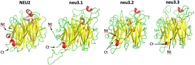
Fig. 5 Ribbon diagram of Hs NEU2 and Dr Neu3.1, 2 and 3 viewed by the side. The active site is located on the top part of the proteins; NEU2 structure (left) shows the competitive inhibitor DANA (violet), as well as the side chain of Asp 46 and Glu 111 (red) located in the mobile loops bearing α1 and α2 helices (PDB code 1VCU). The 3D structural features of the Dr Neus obtained by molecular modelling show a more complex network of unordered segments. The amino and carboxy terminus are indicated by Nt and Ct, respectively. Color code: β-strands yellow; α-helix red; unordered green.
Acknowledgments
This image is the copyrighted work of the attributed author or publisher, and
ZFIN has permission only to display this image to its users.
Additional permissions should be obtained from the applicable author or publisher of the image.
Full text @ Biochimie