
|
Fig. 2.
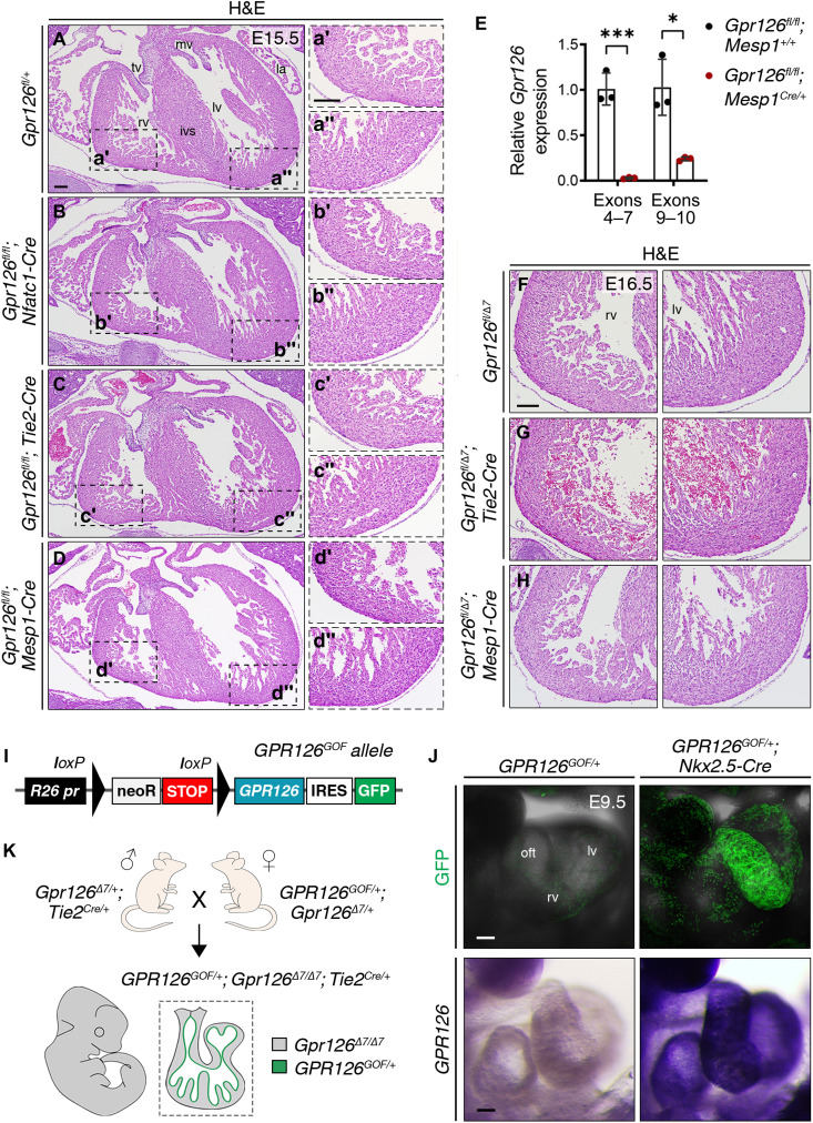
(A to D″) H&E staining of transverse heart sections from WT (A to A″), Gpr126fl/fl;Nfatc1Cre/+ (B to B″), Gpr126fl/fl;Tie2Cre/+ (C to C″), and Gpr126fl/fl;Mesp1Cre/+ (D to D″) embryos at E15.5. Magnified views of the right ventricle are shown in (′) and of the left ventricle in (″). (E) qRT-PCR analysis showing relative Gpr126 gene expression (to Gapdh) in Gpr126fl/fl;Mesp1+/+ and Gpr126fl/fl;Mesp1Cre/+ embryonic hearts at E12.5 using primers spanning exons 4 to 7 and exons 9 and 10. Data are means ± SD (n = 3 embryonic hearts); t test, *P < 0.05 and ***P < 0.001. (F to H) H&E staining of transverse heart sections from WT Gpr126fl/Δ7 (F), Gpr126fl/Δ7;Tie2Cre/+ (G), and Gpr126fl/Δ7;Mesp1Cre/+ (H) embryos at E16.5. Left panels: Right ventricle; Right panels: Left ventricle. (I) Schematic of the conditional gain-of-function R26-GPR126GOF construct. GPR126 expression is activated upon the Cre-mediated excision of the NeoR-STOP cassette. (J) Top panels: Confocal images showing eGFP expression in E9.5 GPR126GOF/+ and GPR126GOF/+;Nkx2.5Cre/+ hearts. Bottom panels: WISH showing GPR126 expression in E9.5 GPR126GOF/+ and GPR126GOF/+;Nkx2.5Cre/+ hearts. (K) Representation of GPR126GOF/+;Gpr126Δ7/Δ7;Tie2Cre/+ embryos from crosses of Gpr126Δ7/+;Tie2Cre/+ males with GPR126GOF/+;Gpr126Δ7/+ females. Scale bars, 100 μm. GFP, green fluorescent protein; IRES, internal ribosome entry site; mv, mitral valve; neoR, neomycin resistance gene; R26 pr, Rosa26 promoter; tv, tricuspid valve.