Figure 2
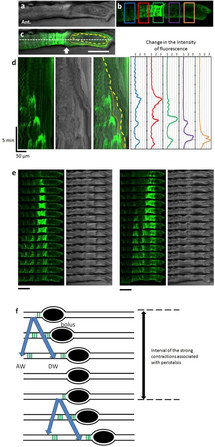
Peristaltic reflex visualized by Ca2+ imaging of circular smooth muscles. (a–c) Live confocal image of the lateral view of SAGFF(LF)134A; Tg(UAS: GCaMP3) at 8 dpf. Bright field image (a), GCaMP3 fluorescence (green) (b), and merged image (c). In (b), measurement locations are shown in rectangles. In (c), the yellow dashed line represents the outline of the bolus. The yellow dotted line represents the outline of the liquid surrounding the bolus. Scale bar, 50 μm. (d) Kymograph of GCaMP3 fluorescence, bright field image and merged image at the site noted by the white dashed line in (c), and time course of the fluorescence intensity at each site noted by the five squares in (b). (e) Time-lapse series of the second (left) and third (right) events in (d). Scale bars, 50 μm. Interval, 11 s. (f) Schematic diagrams showing Ca2+ events in the circular muscles at the peristaltic movement. Black ellipses denote bolus. Also see Supplementary video 1.

