
|
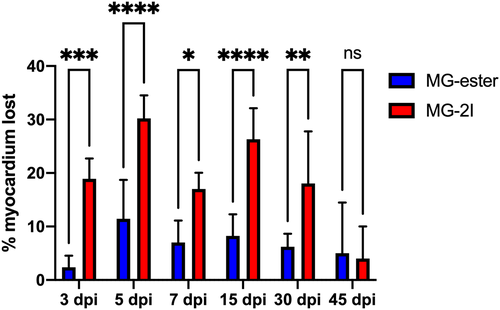
Fig. 2 Chemoptogenetic damage causes loss of myocardium after MG-2I treatment. Regions of Interest were taken from WT uninjured, Tg(myl7:fapdl1-cerulean) MG-ester-, and Tg(myl7:fapdl1-cerulean) MG-2I-treated hearts after AFOG staining and intact myocardium was measured using Threshold Particle Analysis (Image J). Myocardial area from MG-ester and MG-2I hearts was normalized to WT uninjured hearts to generate the amount of tissue lost as a percentage. MG-2I-treated hearts showed significant loss of myocardium at 3 dpi and 5 dpi (compared to MG-ester controls), and also regenerated this damage by 45-60dpi. A minimum of three hearts were used per condition with the following as the total n values: WT uninjured (n = 6), MG-ester (3 dpi (n = 5), −5 dpi (n = 5), −7 dpi (n = 5), −15 dpi (n = 5), −30 dpi (n = 5), −45 dpi (n = 3)), and MG-2I (3 dpi (n = 5), −5 dpi (n = 5), −7 dpi (n = 6), −15 dpi (n = 6), −30 dpi (n = 5), −45 dpi (n = 3)). *P < .05; **P < .01; ***P < .001; ****P < .0001; Two-way ANOVA using Šidák's multiple comparisons test. Values without asterisks are not statistically different from MG-ester control samples