
|
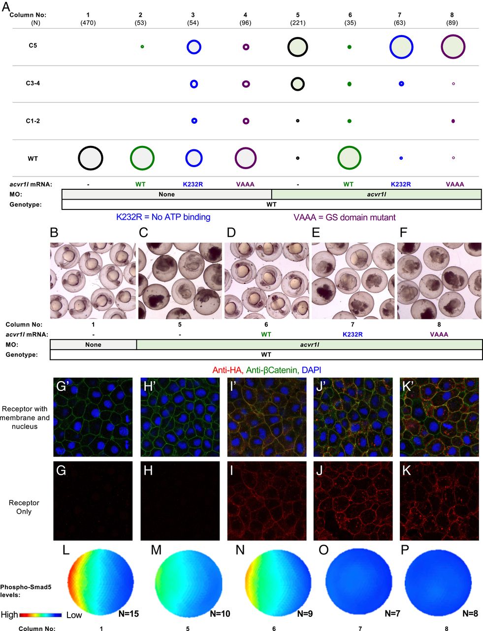
Fig. 4 Acvr1l kinase function is essential for BMP signaling. (A) Kinase-dead Acvr1l cannot signal and restore DV patterning. WT embryos injected with acvr1l-HA RNA (100 pg), kinase-dead acvr1l-K232R-HA RNA (60 pg), and GS-domain mutant acvr1VAAA-HA RNA (100 pg), with or without acvr1l MO. MO concentrations and combinations are listed in SI Appendix, Table S4. Raw phenotype scores are in Dataset S3. (B–F) The acvr1l MO-injected embryos at ∼24 hpf. Injection conditions and corresponding columns in A are labeled below the images. Uninjected embryos are WT (B) Acvr1l-deficient C5 embryos lyse by 24 hpf (C). (D) WT acvr1l-HA RNA fully rescues Acvr1l-deficient embryos. (E and F) Acvr1l-deficient embryos injected with (E) acvr1l-K232R-HA RNA or (F) acvr1l-VAAA-HA RNA remain C5 dorsalized and lyse by 24 hpf. (G–K) Immunostaining against HA-tagged Acvr1l receptor with injection conditions labeled above the images. Embryos ≥3 were imaged for each condition. G’–K’ are the same images as G–K but showing DAPI-stained nuclei (blue) and membrane-localized β-catenin (green). (L–P) Quantified and averaged phospho-Smad5 immunostained embryos from various Acvr1l injection conditions. Corresponding columns in A are labeled on the Bottom. (L) Uninjected embryos display a WT phospho-Smad5 gradient. (M) Acvr1l-deficient embryos display reduced phospho-Smad5. (N) WT acvr1l RNA restores phospho-Smad5 signaling in Acvr1l-deficient embryos, while acvr1l-K232R RNA (O) and acvr1l-VAAA RNA (P) do not.