
|
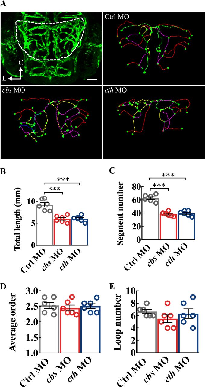
Fig. 3 Structure analysis of the midbrain vasculature in cbs and cth morphants. (A) Image of a 3-dpf larva showing the midbrain position delineated with dashed lines (upper left), and representative midbrain vasculature centerlines of 3-dpf larvae of Ctrl morpholino oligonucleotide (MO; upper right), cbs MO (lower left), and cth MO (lower right). C, caudal; L, lateral. (B–E) Summary of changes in the total vessel length (B), vessel segment number (C), weighted average segment Strahler order (D) and internal vessel loop number (E) of the midbrain vasculature in groups of Ctrl MO, cbs MO and cth MO. Six embryos were examined for each group. Error bars, SEM. ***P<0.001 (unpaired two-tailed Student’s t-test).