
|
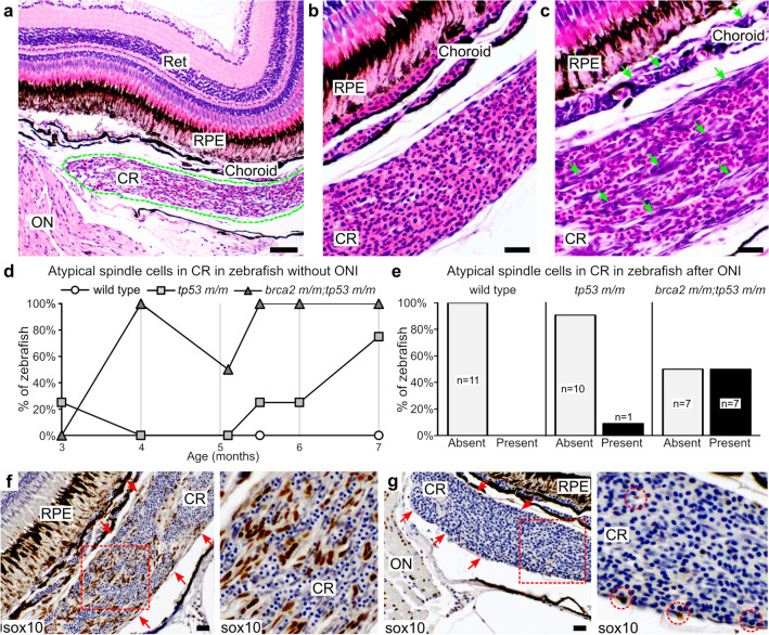
Figure 2
Atypical spindle cells accumulate in the choroid rete of brca2 m/m;tp53 m/m zebrafish. (a) The choroid rete (outlined in green) is a vascular plexus located subjacent to the choroid. (b) Normal choroid rete. (c) Choriod rete containing numerous atypical spindle cells (green arrows). (d) Numbers of zebrafish that developed atypical spindle cells in the choroid rete over time (n = 4 per genotype at each time point analyzed). (e) Numbers of zebrafish that received optic nerve injury and developed atypical spindle cells in the choroid rete. (f) Atypical spindle cells in the choroid rete are sox10-positive (brown chromogen). Red arrows delineate margins of choroid rete. Area boxed in red is shown at higher magnification to the right. (g) Small numbers of sox10-positive cells (brown chromogen) are present in the normal choroid rete. Red arrows delineate margins of choroid rete. Area boxed in red is shown at higher magnification to the right. Red circles identify sox10-positive cells. Ret, retina; RPE, retinal pigmented epithelium; CR, choroid rete; ON, optic nerve. Scale bar = 50 µm (panel a); 20 µm (panels b, c, f, h).