
Figure 3
- ID
- ZDB-IMAGE-220131-188
- Publication
- Ahuja et al., 2022 - Myocardial Afterload Is a Key Biomechanical Regulator of Atrioventricular Myocyte Differentiation in Zebrafish
- All Figures
- Figures for Ahuja et al., 2022
|
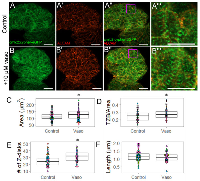
Figure 3 Increased pressure causes hypertrophy of ventricular outer curvature (OC) myocytes by 56 hpf. (A) Control and (B) vaso-treated hearts showing z-disks (A,B) and cell shape through ALCAM immunohistochemistry (A′,B′). (A′′,B′′) display merged images. Purple box shows regions of interest shown in (A′′′,B′′′). For A–A′′, and B–B′′, scale bar represents 20 µm. (A′′′) Control and (B′′′) vaso-treated hearts. Scale bar represents 10 µm. (C) Area of ventricular OC cells in control and vaso-treated hearts. (D) Total z-disk size (TZB) of sarcomeres present in each cell normalized to the area of each cell (1/µm). For details for how TML was measured, see supplemental Figure S11. (E) Number of z-disks per cell in control and vaso-treated hearts. (F) Average length of the sarcomeres present in each cell. For control and vaso-treated hearts, 14 fish were analyzed. Five cells from the OC were analyzed per fish, such that 70 total cells were analyzed. Each colored dot on the graph above represents an individual fish. * indicates p < 0.05, Student’s t-test was used for statistical comparisons