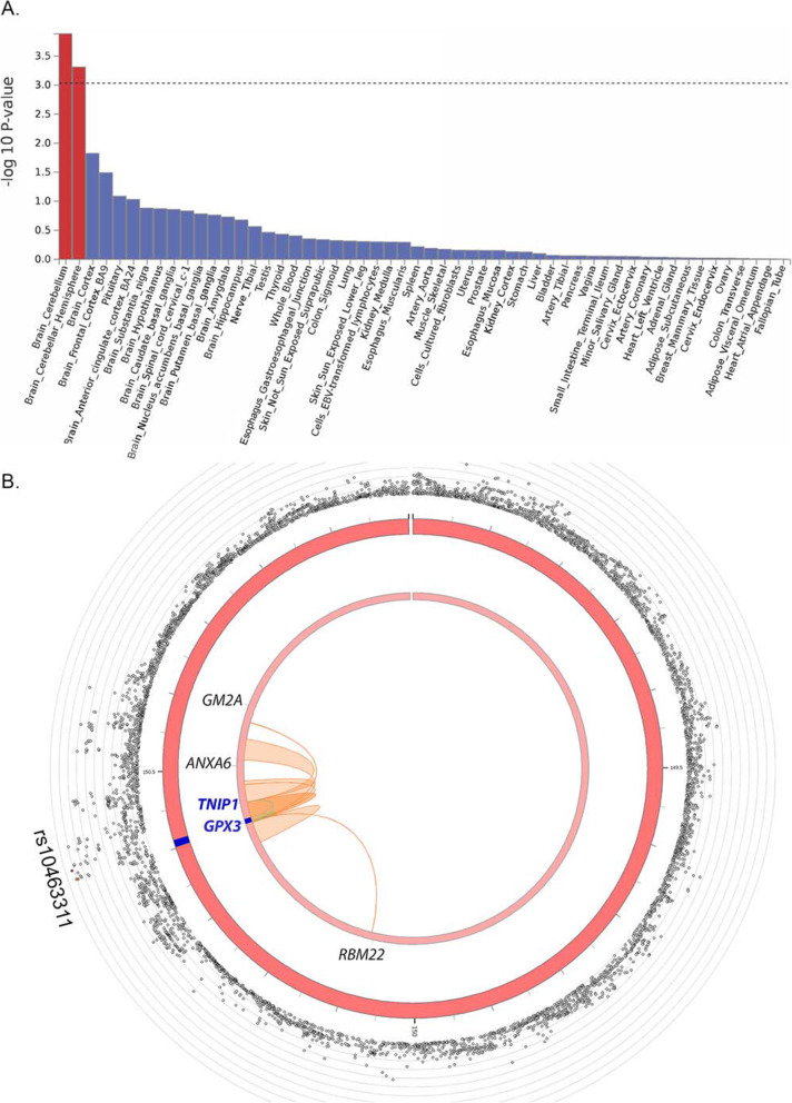
Fig. 2
Functional annotation of ALS GWAS. A Using the full GWAS summary data, enrichment of association signal is identified in genes expressed in brain tissue using GTEx v8 (n = 54 tissues). B Circos plot of chromosome 5 with the risk locus in blue (middle circle); outer circle shows SNP associations (grey circles) with -log10(p-value) on the Y-axis. The lead SNP (rs10463311) is labelled, and other SNPs are coloured if they are in LD of the lead SNP (yellow to red, low to high r2, see Fig. Fig.33 for detail). Inner circle: The mapped genes are labelled black if chromatin interaction is detected (GM2A, ANXA6, RBM22), and blue if both a chromatin interaction and an eQTL is detected (TNIP1, GPX3)

