
|
Figure 4
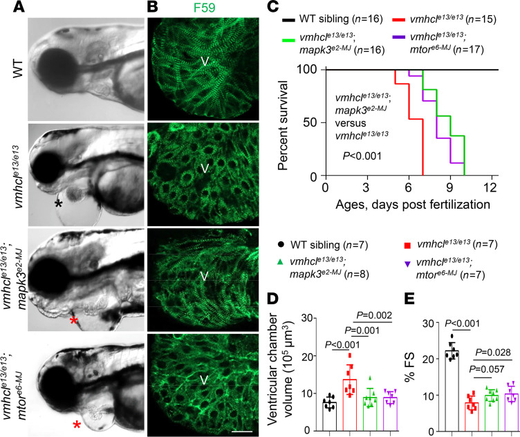
(A) Representative images of the heart area of F0 fish at 3 dpf. The black star indicates severe edema, and red stars indicate mild edema. Scale bar: 300 μm. (B) Fluorescence immunostaining images using anti-myosin heavy chain 1 (F59) in the ventricles of (from top to bottom): WT controls, vmhcle13/e13, vmhcle13/e13;mapk3e2-MJ, and vmhcle13/e13;mtore6-MJ mutant hearts at 3 dpf. Scale bar: 2 μm; V, ventricle. (C) Kaplan–Meier survival curves of vmhcle13/e13 mutant fish upon mapk3 and mtor inhibition and WT controls. n = 15–17; log-rank test. (D and E) VCV (D) and percent FS (E) of the vmhcle13/e13 mutants after mapk3 and mtor inhibition compared to WT controls at 3 dpf. n = 7–8; data are presented as the mean ± SD; 1-way ANOVA.