
Fig. 3
- ID
- ZDB-IMAGE-220124-1
- Antibodies
- Publication
- Taler et al., 2019 - Lysyl hydroxylase 3 is required for normal lens capsule formation and maintenance of lens epithelium integrity and fate
- All Figures
- Figures for Taler et al., 2019
|
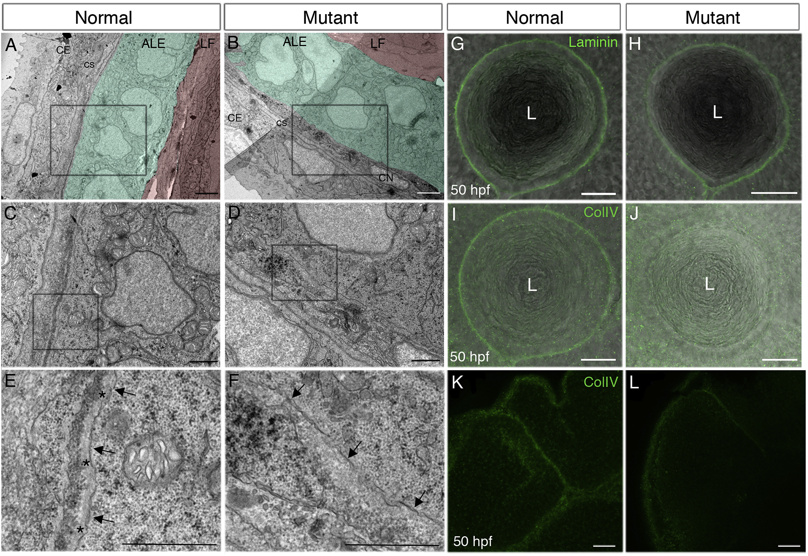
Fig. 3 Lens capsule is abnormal in pninavu222 mutants. (A–F) Representative transmission electron microscope (TEM) images of lens epithelium and lens capsule in a normal sibling (A,C,E) and pninavu222/vu222 (B,D,F) embryo at 48 hpf (n = 3 embryos for each genotype). (A,B) The color coding highlights lens fibers (LF) (red), anterior lens epithelium (ALE) (green) and the bi-layered corneal epithelium (CE) (no color). Corneal stroma (CS) and corneal endothelium (CN) are at early stages of formation. C and D are higher magnifications of the regions marked by rectangles in A and B, respectively. E and F are higher magnifications of the regions marked by rectangles in C and D, respectively. Arrows in E, F point at basal cell membranes of anterior LECs and asterisks in E mark the lens capsule. (G–L) Single confocal planes from 50 hpf normal (G,I,K) and mutant (H,J,L) embryos, lateral views, anterior to the left. Labeling (green) is for Laminin (G,H) or ColIV (I–L). Images show the equator region of lenses (G–J) or forebrain and midbrain regions (K,L). Bright-field images in G-J are overlaid to highlight tissue morphology. Numbers of embryos from two repetitions for each staining: G = 7, H = 9, I = 5, J = 7. L, lens. Scale bars: A,B - 2 μm; C–F - 1 μm; G-L - 20 μm.
Reprinted from Developmental Biology, 458(2), Taler, K., Weiss, O., Rotem, S., Rubinstein, A.M., Seritrakul, P., Gross, J.M., Inbal, A., Lysyl hydroxylase 3 is required for normal lens capsule formation and maintenance of lens epithelium integrity and fate, 177-188, Copyright (2019) with permission from Elsevier. Full text @ Dev. Biol.