
|
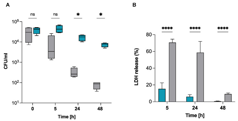
Figure 3
Survival capacity of S. pettenkoferi SP165 in non-professional phagocytes. (A) At a MOI of 100, bacteria from S. pettenkoferi SP165 (blue) and S. aureus SA564 (grey) were utilized to infect cells of the keratinocyte cell line HaCaT, and the cells were co-cultured for 90 min. Washing and lysostaphin/gentamicin treatment were used to eliminate extracellular and adhering bacteria, and infected cells were grown for up to 48 h in cell culture media supplemented with lysostaphin. Eukaryotic cells were lysed, and surviving bacteria in lysates were evaluated by counting CFUs at 5, 24, and 48 h after lysostaphin/gentamicin treatment. Percentage survival = (#CFUfinal/#CFUinput) ∗ 100. (B) LDH release was measured using the CyQUANT assay kit after HaCaT cells were infected at a MOI of 100 for 5, 24 and 48 h, with the exception that the cells were seeded in a 96-well plate, and the extracellular bacteria were not eliminated. Data are expressed relative to the 100% positive control. Statistical significance was determined by two-way ANOVA test where ns, non-significant; * p < 0.05; **** p < 0.0001. n = 4 biological repeats.