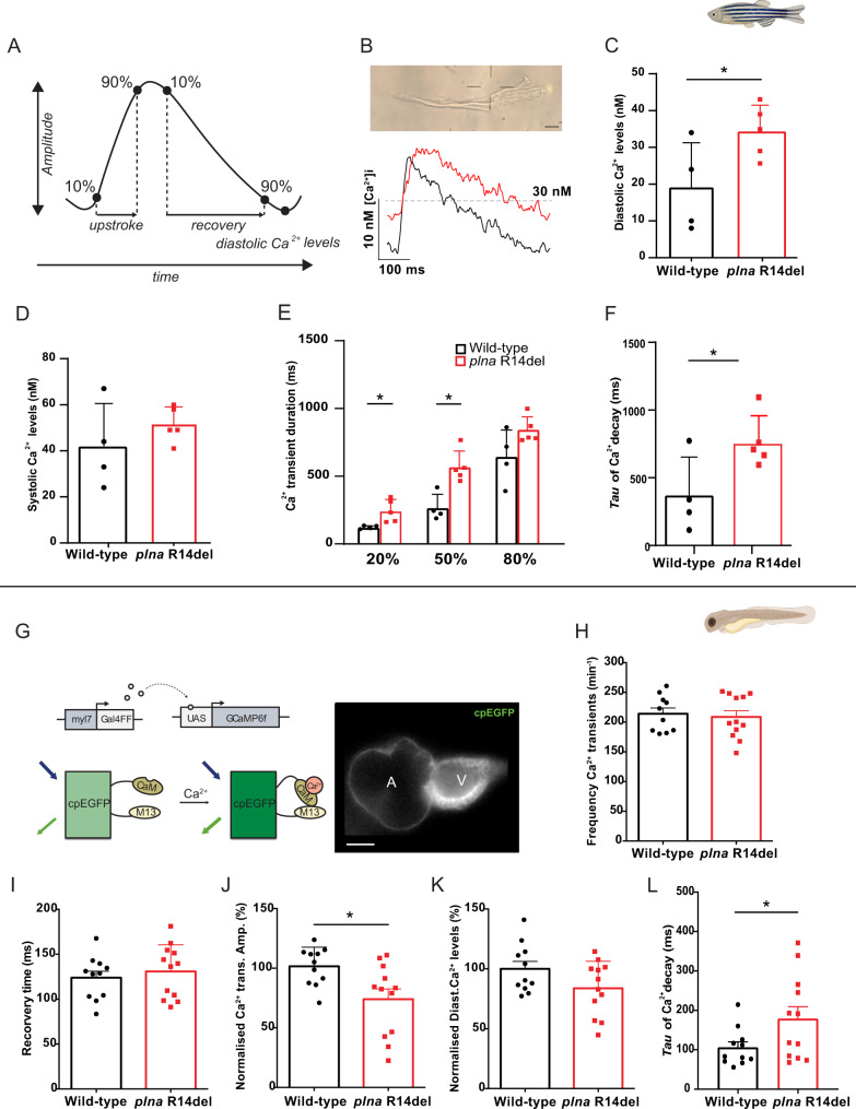
Fig. 5 A Schematic representation of Ca2+ transient parameters showing Ca2+ transient upstroke time, Ca2+ transient recovery time, Ca2+ transient amplitude, and diastolic Ca2+ levels. B Representative image of an isolated ventricular cardiomyocyte and a Ca2+ trace from adult ventricular cardiomyocyte of wild type and plna R14del mutants. Intracellular Ca2+ parameters were measured in adult ventricular cardiomyocytes of wild type (WT) and plna R14del, including C diastolic Ca2+ levels (mean ± SEM, *p = 0.05, WT n = 4, plna R14del n = 5), D systolic Ca2+ levels, E Calcium transient duration at 20%, 50, and 80% (mean ± SEM, CaD20: p = 0.026 and CaD50: p = 0.005, WT n = 4, plna R14del n = 5), and F Tau of Ca2+ decay (mean ± SEM, *p = 0.041, WT n = 4, plna R14del n = 5). All measurements were performed in three experimental replicates. G DNA construct and sensor dynamics of GCaMP6f (left panel). GCaMP6f was placed under the control of the myl7 promoter to restrict its expression to the heart. The Gal4FF-UAS system amplifies its expression. GCaMP6f consists of a circularly permutated enhanced green fluorescence protein (cpEGFP) fused to calmodulin (CaM) and the M13 peptide. When intracellular calcium (Ca2+) rises, CaM binds to M13, causing increased brightness of cpEGFP. Using a high-speed epifluorescence microscope, movies of 3 dpf non-contracting GCaMP6f embryonic hearts were recorded. Several intracellular Ca2+ parameters were measured, including H Frequency of Ca2+ transients, I Ca2+ transient recovery time, J normalized Ca2+ transient amplitude (mean ± SEM, *p = 0.0129, WT n = 10, plna R14del n = 12), K normalized diastolic Ca2+ levels, and L Tau of Ca2+ decay in wild-type (WT) and plna R14del embryonic zebrafish (mean ± SEM, *p = 0.0498, WT n = 10, plna R14del n = 12). All measurements were performed in three experimental replicates. Wild type is highlighted in black and plna R14del in red. Statistical test: unpaired Student’s t-test. nM nanomolar, ms milliseconds, CaM calmodulin, UAS upstream activation sequence, Ca2+ calcium. Source data are provided as a Source Data file.
Image
Figure Caption
Acknowledgments
This image is the copyrighted work of the attributed author or publisher, and
ZFIN has permission only to display this image to its users.
Additional permissions should be obtained from the applicable author or publisher of the image.
Full text @ Nat. Commun.

