
|
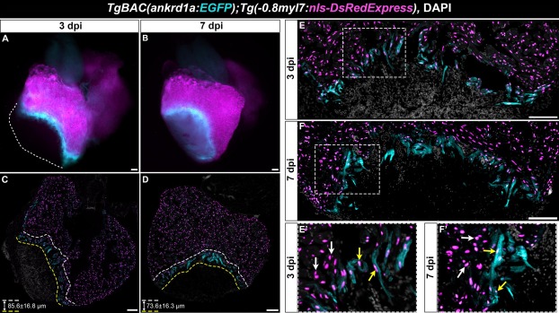
Fig. 5 TgBAC(ankrd1a:EGFP) is highly expressed in cardiomyocytes at the cryoinjury border zone. Hearts of TgBAC(ankrd1a:EGFP);Tg(-0.8myl7:nls-DsRedExpress) zebrafish were injured and left to recover for 3 and 7 days. (A, B) Images of whole hearts reveal localization of EGFP+ positive cells at the border with injured tissue. (C, D) Cryosections of injured hearts at 3 and 7 dpci. Yellow and white dotted lines outline the zones of EGFP+ cells. Average thickness ± SD of these zones is displayed in the lower left corner of each panel. (E, F) Higher magnification of injury border zone at 3 and 7 dpci. (E’, F’) Higher magnification of areas from E and F, showing injury border zone CMs. Yellow arrows point to EGFP+/nls-DsRedExpress+ and white arrows point to EGFP-/nls-DsRedExpress+ CMs. Scale bars, 100 µm. (For interpretation of the references to color in this figure legend, the reader is referred to the web version of this article.)
Reprinted from Gene, 792, Boskovic, S., Marin Juez, R., Stamenkovic, N., Radojkovic, D., Yr Stainier, D., Kojic, S., The stress responsive gene ankrd1a is dynamically regulated during skeletal muscle development and upregulated following cardiac injury in border zone cardiomyocytes in adult zebrafish, 145725, Copyright (2021) with permission from Elsevier. Full text @ Gene