
Image
|
Figure Caption
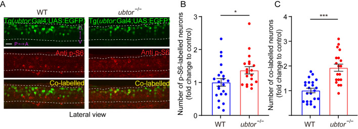
Fig. 4
mTORC1 hyperactivation in the spinal interneurons of ubtor mutant embryos. A p-S6 staining and EGFP signals in 28-hpf embryos (dotted lines outline spinal somites 4–10; scale bar, 40 μm). B Numbers of p-S6-labelled neurons in the spinal cord of 28-hpf embryos (three biological repeats, NWT = 22, Nubtor−/− = 19, t39 = 5.538). C Numbers of co-labeled p-S6/EGFP neurons in the spinal cord of 28-hpf embryos (three biological repeats, NWT = 22, Nubtor−/− = 19, t39 = 2.265). Values are represented as the mean ± SEM in B and C. *P <0.05, ***P <0.001.
Acknowledgments
This image is the copyrighted work of the attributed author or publisher, and
ZFIN has permission only to display this image to its users.
Additional permissions should be obtained from the applicable author or publisher of the image.
Full text @ Neurosci. Bull.