
|
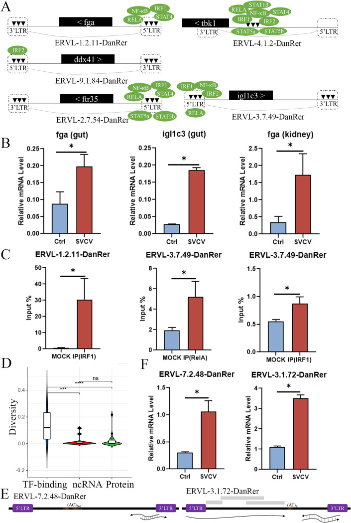
FIG 9
Potential regulatory elements and transcriptional expression analysis of DrERVs upon SVCV infection. (A) Schematic diagram of the potential TF-binding sites at the LTRs of DrERVs inserted with different functional genes that were actively expressed in response to SVCV stimulation. These DrERVs are designated as virus-responsive ERVs (VREs), and their associated functional genes are named as VRE-aid genes. The positional relationship between VREs and VRE-aid genes is shown. (B) Transcriptional expression analysis of two representative VRE-aid genes in head kidney and gut tissues upon SVCV infection. (C) Examination of the TF-binding activity of two representative TFs (IRF1 and RelA) at the LTRs of two VREs by ChIP–qPCR analysis. (D) Comparison of the sequence diversity of LTRs among different VRE types. (E) Schematic diagram of two typical noncoding VREs and the transcripts. The gray box represents the additional LTRs detected inside the DrERVs. (F) Expression analysis of noncoding VREs upon SVCV stimulation (*, P < 0.05; ***, P < 0.001; ****, P < 0.0001; ns, no significant difference).