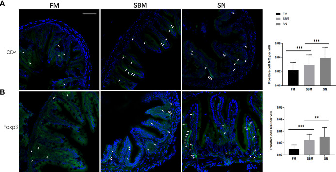
Lymphocyte-related intestinal protein expression reflected by immunofluorescent signals. CD4+(A) or Foxp3+(B) immunofluorescence signals (green) in adult fish fed with FM, SBM, and SN diets. The quantification of the ratio, which was calculated by dividing positive cell number with intestinal villi’s length, was shown beside the typical images (at the right side). Typical signals are indicated by arrows. The blue signals represented DAPI-stained cell nucleus. FM, fish meal diet; SBM, soybean meal diet; SN, sinomenine supplementary SBM diet. Scale bar: 100 μm. ** represented p < 0.01, and *** represented p < 0.001.
Acknowledgments
This image is the copyrighted work of the attributed author or publisher, and
ZFIN has permission only to display this image to its users.
Additional permissions should be obtained from the applicable author or publisher of the image.
Full text @ Front Immunol
Your Input Welcome
Thank you for submitting comments. Your input has been emailed to ZFIN curators who may contact you if
additional information is required.
Oops. Something went wrong. Please try again later.