
Fig. 6
- ID
- ZDB-IMAGE-211203-16
- Genes
- Publication
- Cavone et al., 2021 - A unique macrophage subpopulation signals directly to progenitor cells to promote regenerative neurogenesis in the zebrafish spinal cord
- All Figures
- Figures for Cavone et al., 2021
|
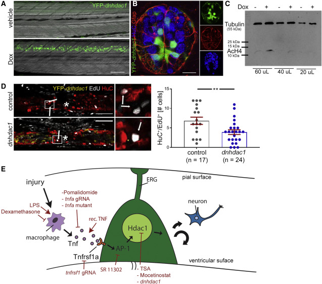
Fig. 6 Hdac1 function in ERG progenitor cells is necessary for regenerative neurogenesis (A) Doxycycline (Dox) exposure leads to strong detectability of YFP in tg(her4.3:TetA; TetRE:YFP-dnhdac1) transgenic animals at 24 h post-induction; lateral views are shown (rostral left; dorsal up). (B) A cross section (dorsal up) through the spinal cord of a tg(her4.3:TetA; TetRE:YFP-dnhdac1) larva shows labeling in ventricular ERG progenitors, some of which are glial fibrillary acidic protein (Gfap) immune positive. Peripheral HuC immuno-labeled neurons are rarely labeled by the trasngene. (C) Western blot shows a doxycycline-induced increase in histone acetylation levels (AcH4) in FACS purified ERGs. Anti-tubulin labeling was used as a standard. (D) Activation of transgene expression reduces the number of newly generated neurons (HuC+/EdU+, t test: ∗∗p = 0.009; incubation time for doxycycline and EdU: 0 to 48 hpl). Control is doxycycline-treated non-transgenic siblings. (E) Graphical summary of immune system mediated regenerative neurogenesis with experimental interventions indicated in red. Scale bars: 50 μm. Data are represented as mean ± SEM.
Reprinted from Developmental Cell, 56(11), Cavone, L., McCann, T., Drake, L.K., Aguzzi, E.A., Oprişoreanu, A.M., Pedersen, E., Sandi, S., Selvarajah, J., Tsarouchas, T.M., Wehner, D., Keatinge, M., Mysiak, K.S., Henderson, B.E.P., Dobie, R., Henderson, N.C., Becker, T., Becker, C.G., A unique macrophage subpopulation signals directly to progenitor cells to promote regenerative neurogenesis in the zebrafish spinal cord, 1617-1630.e6, Copyright (2021) with permission from Elsevier. Full text @ Dev. Cell