IMAGE

Figure 1

ID
ZDB-IMAGE-211201-12
Source
Figures for Siegerist et al., 2021
Image
Figure Caption

Figure 1

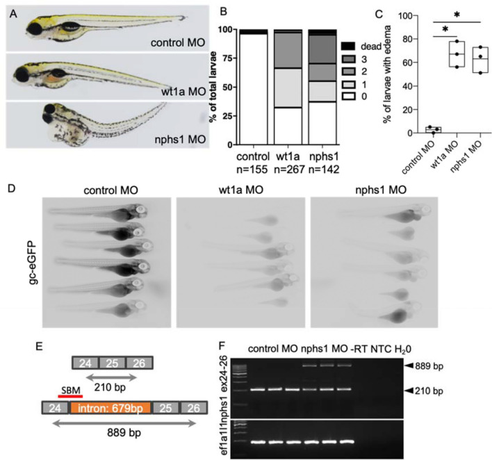
Induction of primary glomerular disease in larval zebrafish: Zebrafish embryos were injected with 2 nl 100 µM translation-blocking anti wt1a, splice-blocking anti-nphs1 or non-binding control MOs. (A) Total of n = 564 zebrafish embryos were injected and phenotyped at 120 hpf. Phenotype distribution graded in 0:no edema to 3: severe edema with bent body axis and dead larvae. As shown in the stacked boxplots in (B), the phenotype of the wt1a knockdown larvae was generally more severe compared to nphs1 knockdown while the general proportion of larvae with edema was similar in both groups (C). No overt edema was seen in control MO injected larvae and the percentage of larvae with edema of any kind was statistically significant higher compared to paired control groups (paired t-test, p = 0.01). Shown in panel (D) are the results of the zebrafish proteinuria assay. While control MO-injected Tg(fabp10a:gc-eGFP) larvae showed intravascular localization of gc-eGFP, wt1a and nphs1 MO-injected larvae showed significantly less eGFP-fluorescence indicating clearance of the 78 kDa fusion protein from the blood plasma. (E) shows the exon–intron structure of exons 24–26 in the nphs1 gene with respective PCR-primer sites and the SBM MO binding site. Binding of the nphs1 SBM should lead to integration of a 679 bp intron. (F) shows agarose gel-resolved RT-PCR products for the nphs1 region described in (D) for three independent control and nphs1 SBM injected groups. Arrowheads show the size of the respective wildtype-bands at 210 and the shifted PCR product after intronic integration.

Acknowledgments
This image is the copyrighted work of the attributed author or publisher, and ZFIN has permission only to display this image to its users. Additional permissions should be obtained from the applicable author or publisher of the image. Full text @ Sci. Rep.