IMAGE

Fig. 4

ID
ZDB-IMAGE-211129-14
Source
Figures for Lin et al., 2021
Image
Figure Caption

Fig. 4

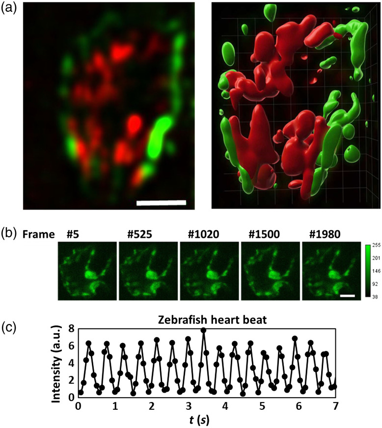
Two-color 2p LSFM with axicon imaging of a zebrafish heart beating. (a) A slice of a volume image and its 3D rendering image (10 slices; 100𝜇m×100𝜇m×20𝜇m) recorded at 10 volumes per second. The transgenic zebrafish Tg(fli1a:EGFP; gata1:DsRed) expressed EGFP in the vascular endothelium and Dsred in RBCs. (b) Representative time-lapse images of a zebrafish heart and (c) zebrafish beating heart dynamics. EGFP fluorescence signal was recorded within an area of the heart as a function of time. The measured heartbeat was 2.2 Hz at 20°C. Scale bar: 30𝜇m.

Acknowledgments
This image is the copyrighted work of the attributed author or publisher, and ZFIN has permission only to display this image to its users. Additional permissions should be obtained from the applicable author or publisher of the image. Full text @ J. Biomed. Opt.