Figure 3
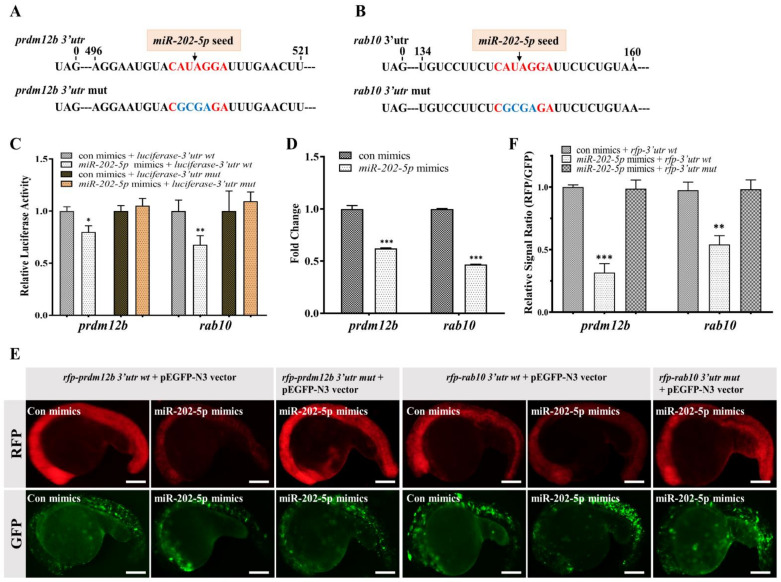
miR-202-5p directly regulates prdm12b and rab10. (A,B) The sequence information of the 3′utr of prdm12b and rab10. The canonical miR-202-5p binding sites and its mutant were shown in red and blue, respectively. (C) Relative luciferase activity in HEK293T cells transfected with psiCHECK2-3′utr wt (prdm12b or rab10) or psiCHECK2-3′utr mut (prdm12b or rab10) in the presence of control mimics (con mimics) or miR-202-5p mimics. (D) qRT-PCR analysis of prdm12b and rab10 mRNA in 24hpf embryos injected with control or miR-202-5p mimics. (E) The representative images of embryos co-injected with mRNAs of rfp-prdm12b 3′utr wt, rfp-prdm12b 3′utr mut, rfp-rab10 3′utr wt or rfp-rab10 3′utr mut in the presence of control or miR-202-5p mimics. All embryos were co-injected with pEGFP-N3 vector as a control. (F) The relative signal ratio of RFP/GFP in the experiment presented in (E). (C,D,F) The results were representative of more than three independent experiments in triplicate. β-actin was used as an internal control to normalize gene expression levels with 2-∆∆Ct method * p < 0.05; ** p < 0.01; *** p < 0.001.

