
|
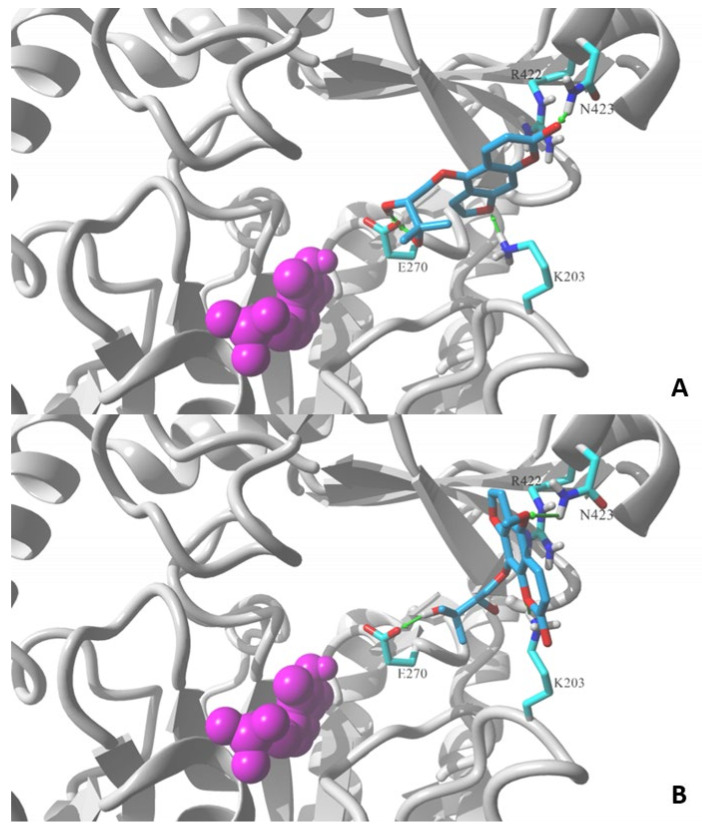
Figure 4
Lowest energy poses of (A) oxypeucedanin hydrate (2) and (B) byacangelicin (8) docked to a structural model of GABA-transaminase cocrystalized with pyridoxal 5′-phosphate (1OHV.pdb). Docked molecules are rendered in stick mode with atom color coded style; protein molecule is rendered in secondary structure mode (gray) and only the residues found essential for interaction with docked molecules are explicitly visualized in the stick model (discussed hydrogen bonds shown as green arrows). Pyridoxal 5′-phosphate cofactor molecule is shown in ball mode and colored in magenta. All aliphatic hydrogen atoms are hidden for clarity. Figures prepared in YASARA 19.5.23.