
|
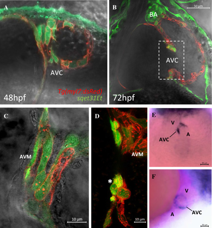
Fig. 1
EGFP expression in the transgenic line sqet31Et defines cells of the AVC. A, B Confocal images showing the AVC region at 48 hpf and 72 hpf. Note the overlap between EGFP and mRFP signals, indicating extensive co-localization and confirming the largely myocardial nature of the EGFP expression domain in sqet31Et. C, D Close-up of the region marked in panel B at different focal planes showing the surface (C) and lumen (D) of the AVC. Note the group of three cuboidal cells protruding into the cardiac lumen at the location corresponding to the developing cardiac cushion (asterisk). E, F Whole-mount in situ hybridization of egfp in sqet31Et transgenic embryos at 72 hpf showing enrichment of EGFP expression in the AVC relative to the rest of the heart. E ventral, D lateral view, A atrium, V ventricle, AVC atrioventricular canal, BA bulbus arteriosus, AVM atrioventricular myocardium