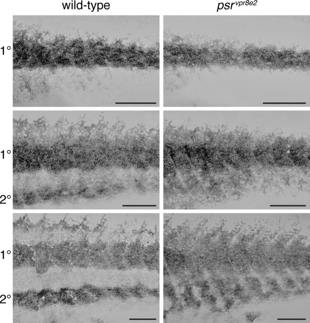
Fig. s3 Development of iridophore pattern in psr mutants. Iridophores were labeled with membrane-targeted mCherry driven by regulatory elements of purine nucleotide phosphorylase 4a (pnp4a:mem-mCherry), which is expressed strongly in these cells (Gur et al., 2020; Spiewak et al., 2018). Pixel values are inverted. Wild-type and psr mutant siblings were imaged repeatedly on successive days through completion of the adult pattern, and two representative individuals are shown here at each of three stages (n = 6 wild-type and 5 psr mutants total). In wild-type, densely packed iridophores formed a primary (1°) interstripe, with more loosely arranged iridophores developing dorsally and ventrally in prospective stripes (top, 9.2 SSL). A secondary (2°) interstripe subsequently formed ventrally (middle, 10.9 SSL) and began to form dorsally (bottom, 13.4 SSL). In psr mutants, densely packed iridophores typical of interstripes were spread more widely over the flank. Scale bars, 500 μM.
Reprinted from Developmental Biology, 476, Eom, D.S., Patterson, L.B., Bostic, R.R., Parichy, D.M., Immunoglobulin superfamily receptor junctional adhesion molecule 3 (Jam3) requirement for melanophore survival and patterning during formation of zebrafish stripes, 314-327, Copyright (2021) with permission from Elsevier. Full text @ Dev. Biol.

