
|
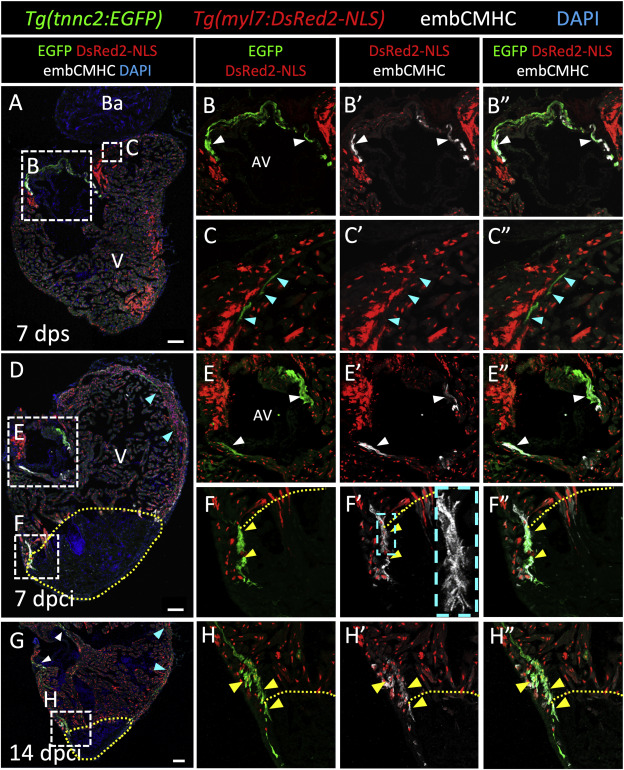
Fig. 6 Cortically located border zone cardiomyocytes upregulate the Tg(tnnc2:EGFP) reporter during adult zebrafish heart regeneration. (A) 7 dps and (D) 7 dpci Tg(tnnc2:EGFP); Tg(myl7:DsRed-NLS) ventricular sections stained for EGFP (green), CM nuclei (red), embCMHC (white) and DNA (blue). (B–C″) Insets show high-magnification images of 7 dps tnnc2:EGFP+/embCMHC+ CMs surrounding the AV valve (B–B″) and tnnc2:EGFP+ primordial layer CMs. (E-F″) Insets show high-magnification images of 7 dpci tnnc2:EGFP+/embCMHC+ CMs surrounding the AV valve (E-E″) and cortically located tnnc2:EGFP+/embCMHC+ CMs protruding into the injured area (F–F″). Cyan dashed box shows a high magnification image of tnnc2:EGFP+/embCMHC+ CMs displaying sarcomeric structures. (G) 14 dpci Tg(tnnc2:EGFP); Tg(myl7:DsRed-NLS) ventricular sections stained for EGFP (green), CM nuclei (red), and DNA (blue). (H–H″) Insets show high-magnification images of 14 dpci cortically located tnnc2:EGFP+/embCMHC+ CMs protruding into the injured area. White arrowheads point to tnnc2:EGFP+ CMs in the AV canal. Cyan arrowheads point to tnnc2:EGFP+ CMs in the primordial layer. Yellow arrowheads point to regenerating tnnc2:EGFP+ CMs in the injured area. V: ventricle; Ba: bulbus arteriosus; AV: atrioventricular canal. Scale bars: 100 μm (A, D, G), 50 μm (B–B″, E-E″), and 20 μm (C–C″, F–F″, H–H″).
Reprinted from Developmental Biology, 476, Tsedeke, A.T., Allanki, S., Gentile, A., Jimenez-Amilburu, V., Rasouli, S.J., Guenther, S., Lai, S.L., Stainier, D.Y.R., Marín-Juez, R., Cardiomyocyte heterogeneity during zebrafish development and regeneration, 259-271, Copyright (2021) with permission from Elsevier. Full text @ Dev. Biol.