
Figure 3
- ID
- ZDB-IMAGE-211103-7
- Publication
- Heylen et al., 2021 - Pericardial Injection of Kainic Acid Induces a Chronic Epileptic State in Larval Zebrafish
- All Figures
- Figures for Heylen et al., 2021
|
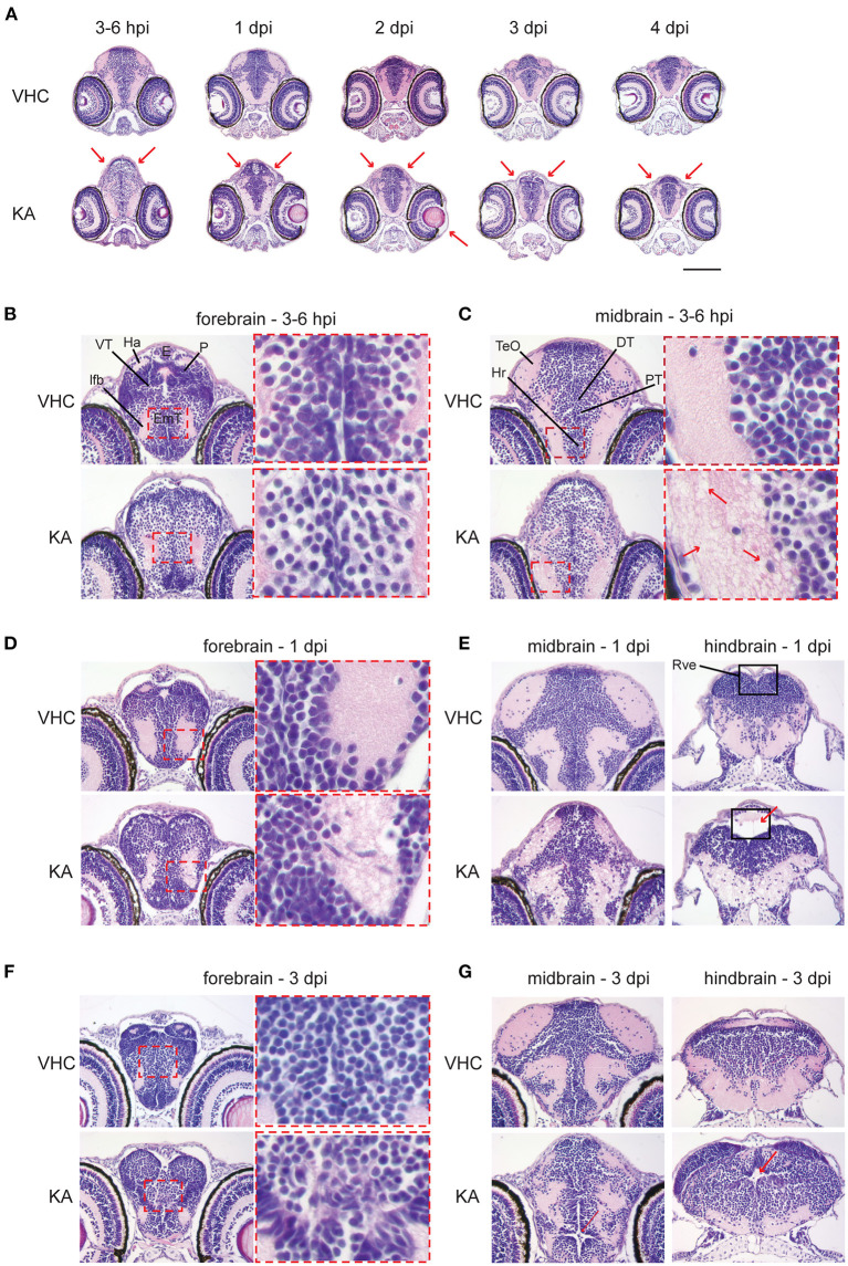
Figure 3
Histopathological analysis reveals that kainic acid (KA) injection induces whole brain abnormalities. Representative images of hematoxylin and eosin (H&E) staining on paraffin cross-brain sections of KA-injected larvae in comparison to VHC-injected siblings. (A) Abnormal brain shape was observed at 2–3 hpi until 4 dpi, marked with red arrows. Scale bar 500 μm. Selected brain abnormalities in KA-injected larvae from different parts of the brain at 3–6 hpi (B,C), 1 dpi (D,E) and 3 dpi (F,G). Red rectangles and arrows highlight the histopathological changes and its magnification. DT, dorsal thalamus; E, epiphysis; EmT, eminentia thalami; Ha, habenula; Hr, rostral hypothalamus; lfb, lateral forebrain bundle; P, pallium; PT, posterior tuberculum; Rve, rhombencephalic ventricle; TeO, optic tectum; VT, ventral thalamus.