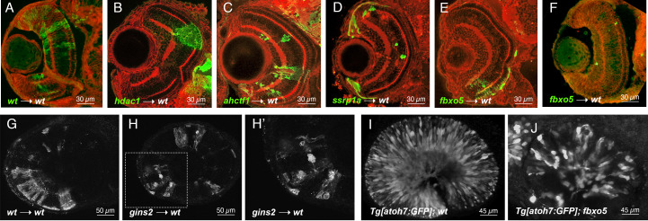
Figure 1
A-F Representative images of frontal cryosections of 3 dpf chimeric wild-type (wt) retinae containing GFP-labelled wild-type (A), hdac1 mutant (B), ahctf1 mutant (C), ssrp1a mutant (D) or fbxo5 mutant (E-F) cells immunostained for GFP (green cells from donor embryo) and beta-catenin (red cell boundaries and plexiform layers).
G-H Lateral views of single 2µm-thick z-plane of whole-mount 3 dpf chimeric wild-type retinae containing wild-type (G) or gins2 morphant (H) cells labeled with membrane-targeted RFP. H’ shows 3x enlargement of boxed region in H.
I-J Lateral maximum intensity projection of ~50 hpf retinae from Tg[atoh7:GFP] embryos showing neurogenic gene expression in wild-type (I) or fbxo5 mutant (J) retinae.

