
Figure 1
- ID
- ZDB-IMAGE-211029-272
- Publication
- Chiang et al., 2021 - Progranulin A Promotes Compensatory Hepatocyte Proliferation via HGF/c-Met Signaling after Partial Hepatectomy in Zebrafish
- All Figures
- Figures for Chiang et al., 2021
|
Figure 1
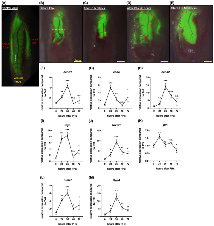
Liver regeneration was induced by one-half ventral lobe partial hepatectomy in 7 days. (A) Ventral view of Tg(fabp10:EGFP). The green fluorescent area marked by the red dotted line is the liver of Tg(fabp10:EGFP). (B) Schematic diagram of one-half ventral lobe partial hepatectomy (PHx). The regenerating ventral lobe is shown at 0 h (C), 96 h (D), and 168 h (E) after PHx. Area marked by the red dotted line is the liver ventral lobe. Yellow dotted line, the site of PHx. Scale bar: 1 mm. Gene expression was examined by quantitative PCR. Cell cycle regulatory genes included (F) ccnd1, (G) ccne, and (H) ccna2. Cell proliferation-associated genes included (I) myc, (J) foxm1, and (K) jun. (L) Gene expression of c-met. (M) Gene expression of GrnA. The relative expression levels compared to control at 0 h PHx are presented as mean ± SD. Significance was set at * p < 0.05, ** p < 0.01, *** p < 0.001, as determined by t-test; PHx, partial hepatectomy.