
|
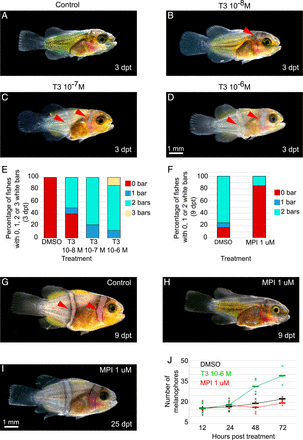
Fig. 3 White bars in A. ocellaris form earlier and later, respectively, after treatments with TH or goitrogens. (A–D) Stereomicroscope images of larvae treated at stage 3 during 3 d (dpt) in DMSO (A) or T3 at 10−6 (B), 10−7 (C), and 10−8 M (D). (E and F) Histogram showing the percentage of larvae having 0 (red), 1 (blue), 2 (turquoise), or 3 (yellow) white bars. (E) Larvae are treated at stage 3 for 3 d with DMSO, T3 10−6, 10−7, and 10−8 M (nDMSO = 16, nT3 10−8 M = 20, nT3 10−7 M = 18, nT3 10−6 M = 15 individuals). χ2 tests are significant between T3 10−6 M and DMSO (P < 0.0001). (F) Larvae are treated at stage 3 for 9 d with DMSO or MPI 1 μM (nDMSO = 12, nMPI 1 μM= 13 individuals). Statistical test was done using χ2 tests (P < 0.0029). (G–I) Stereomicroscope images of larvae treated at stage 3 during 9 d in DMSO (G) and MPI 1 μM (H) and MPI 1 μM stage 3 larvae treated for 25 d (I). (J) Graphic showing the number of melanophores in a specific area of the trunk in DMSO (black), T3 10−6 M (green), and MPI 1 μM (red) at 12, 24, 48, and 72 hpt (nDMSO > 9, nT3 > 9, nMPI > 9 individuals). The statistical tests were done using ANOVA between the T3 or MPI treatments and DMSO (control) at each time. The tests are significant between T3 and DMSO at 48 hpt and 72 hpt (P are respectively equal to 0.0299 and 0.0043). The bars correspond to the mean, and crosses correspond to one experiment. hpt = hours posttreatment (scale bar, 1 mm).