IMAGE

Figure 1

ID
ZDB-IMAGE-211007-19
Source
Figures for Senk et al., 2021
Image
Figure Caption

Figure 1

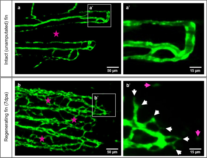
Vascular organization of intact caudal fin vs. early regeneration. Organization of tissue compartments in the intact caudal fin (a) versus the regenerating fin (b), at 7dpa. a/a’ display smooth and hierarchically well-organized blood vessels (green - reporter transgenic zebrafish line) in which intraray segments do not contain any vessels (asterisk) and capillary sprouts are not detectible (a’). (b/b’) display dense, well-interconnected capillary meshwork. The intraray region (purple asterisks) contains bridging vessels and at the expanding vascular front multiple sprouts (b’, white arrows) and migrating - endothelial cells (purple arrows). Images are acquired by fluorescent reflected light microscope.

Acknowledgments
This image is the copyrighted work of the attributed author or publisher, and ZFIN has permission only to display this image to its users. Additional permissions should be obtained from the applicable author or publisher of the image. Full text @ Sci. Rep.