
Figure 7
- ID
- ZDB-IMAGE-211002-153
- Genes
- Publication
- Li et al., 2021 - Functions of SMC2 in the Development of Zebrafish Liver
- All Figures
- Figures for Li et al., 2021
|
Figure 7
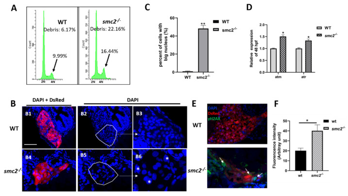
Extensive apoptosis occurring in liver of SMC2−/− mutants is attributable to DNA damage. (A) FACS analyses after DAPI staining of dissociated cells at 96 hpf from wild-type and SMC2−/− embryos. The SMC2−/− embryos have an accumulation of cells in the G2/M phase and more debris. (B) Frozen sections were stained with DAPI to visualize nuclei under the Tg (fabp10a:dsRed; ela3l:EGFP) transgenic background at 96 hpf. The SMC2−/− embryos have an increased number of cells with bigger nucleus compared with WT. Figure 7(B3,B6) are enlargement of the framed part in Figure 7(B2,B5), respectively. *, bigger nuclei. (C) Quantitative analysis of the cell numbers with big nucleus in the liver. Three WT and three mutant embryos across the liver were determined using the ImageJ software. **, p < 0.01. (D) Expression levels of genes (atm and atr) implicated in DNA damage response pathway were analyzed by qPCR in WT and SMC2−/− mutants at 96 hpf. Expression levels were normalized to WT embryos. Data expressed as mean ± SD were representative of three independent experiments. * p < 0.01. (E) Frozen sections were stained against γ-H2AX antibody, which marks DNA double-stranded breaks and nuclei counterstained with DAPI (blue) under the Tg (fabp10a:dsRed; ela3l:EGFP) transgenic background at 96 hpf. The signal was labeled with white arrowhead. (F) Quantitative analysis of the apoptotic cells in the liver. Fluorescence intensities of three WT and three SMC2−/− mutant across the liver were determined using the ImageJ software. **, p < 0.01.