
Figure 3
- ID
- ZDB-IMAGE-210915-52
- Antibodies
- Publication
- Zhu et al., 2021 - Loss of Ift74 Leads to Slow Photoreceptor Degeneration and Ciliogenesis Defects in Zebrafish
- All Figures
- Figures for Zhu et al., 2021
|
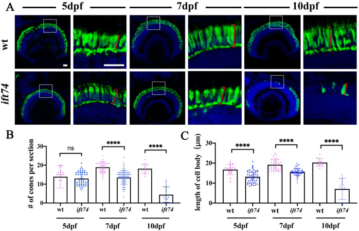
Figure 3
Slow photoreceptor degeneration in ift74 mutants. (A) Confocal images showing the cell bodies of red and green double cones in wild-type and ift74 mutant larvae at different stages as indicated. The double cones were stained with zpr-1 antibody (green), and nuclei were stained with DAPI (blue). Enlarged views of the boxed area are shown on the right. (B,C) Statistical results showing the number of cones per retina section and length of cone cell body in different groups as indicated. Measurements of the cell body length are shown in panel (A) (Red square brackets). Scale bars: 20 µm. **** p < 0.0001, ns, no significant.