
IMAGE
Figure 2
- ID
- ZDB-IMAGE-210915-51
- Antibodies
- Publication
- Zhu et al., 2021 - Loss of Ift74 Leads to Slow Photoreceptor Degeneration and Ciliogenesis Defects in Zebrafish
- All Figures
- Figures for Zhu et al., 2021
Image
|
Figure Caption
Figure 2
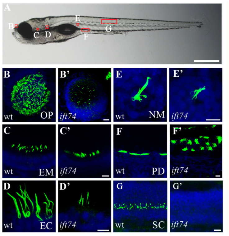
Cilia defects in ift74 mutants. (A) Representative image showing the position of cilia in different organs as indicated in a 5 dpf zebrafish larva. (B–G′) Confocal images showing cilia in different organs of wild-type and ift74 mutants as indicated. Cilia were visualized with anti-glycylated tubulin antibody (green), and nuclei were counterstained with DAPI (blue). OP, olfactory placode; EM, ear macula; EC, ear crista; NM, neuromast; PD pronephric duct; SC, spinal canal. Scale bars: (A), 500 µm; (B′–G′), 10 µm.
Figure Data
Acknowledgments
This image is the copyrighted work of the attributed author or publisher, and
ZFIN has permission only to display this image to its users.
Additional permissions should be obtained from the applicable author or publisher of the image.
Full text @ Int. J. Mol. Sci.