
Figure 3
- ID
- ZDB-IMAGE-210915-33
- Publication
- Berlingerio et al., 2021 - Renal and Extra Renal Manifestations in Adult Zebrafish Model of Cystinosis
- All Figures
- Figures for Berlingerio et al., 2021
|
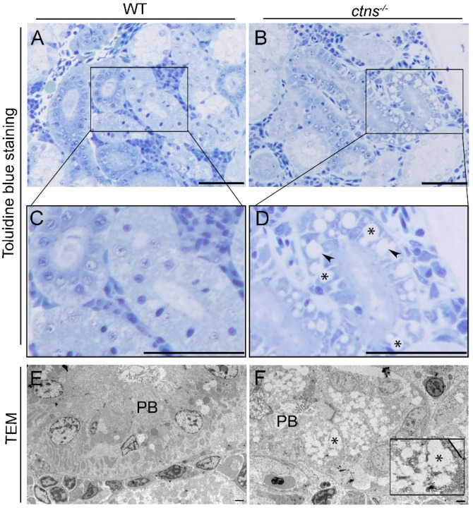
Figure 3
Kidney cystine crystals in wild-type and ctns−/− zebrafish PTECs. (A,B) Representative toluidine blue-stained images of renal proximal tubules in wild-type (A) and ctns−/− (B) 18-month-old zebrafish. The scale bars represent 50 µm. (C,D) Figure 3C,D show a higher magnification of Figure 3A,B. Details of renal proximal tubules with cytoplasmic vacuoles ((D); *), rectangular and polymorphous vacuolar spaces ((D); black arrowheads). (E,F) Representative TEM images of the renal proximal tubule of wild-type (E) and ctns−/− 18-month-old zebrafish (F). (F) Partial loss of proximal tubule brush borders (PB) and accumulation of the rectangular and polymorphous vacuoles (*) were observed in the renal proximal tubule of ctns−/− zebrafish. The high-magnification view of the rectangle in the bottom right corner shows the polymorphous vacuoles and a large vacuole with straight membrane border segments (a straight line). The scale bars represent 5 µm.