
Figure 10
- ID
- ZDB-IMAGE-210915-14
- Publication
- Berlingerio et al., 2021 - Renal and Extra Renal Manifestations in Adult Zebrafish Model of Cystinosis
- All Figures
- Figures for Berlingerio et al., 2021
|
Figure 10
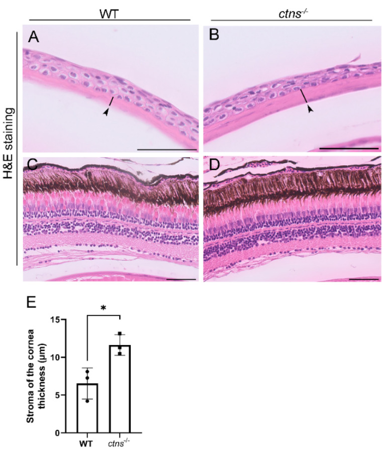
Eye histology in wild-type and ctns−/−zebrafish. (A,B) Representative images of cornea of wild-type (A) and ctns−/−(B) 18-month-old zebrafish. The stromal layer of the cornea (black arrowheads and black lines). H&E staining. The scale bars represent 50 µm. (C,D) Representative images of retina of wild-type (A) and ctns−/−(B) zebrafish. H&E staining. The scale bars represent 50 µm. (E) Relative quantification of the thickness of the stromal layer of the cornea. Each dot represents one zebrafish, for a total of n = 3 wild-type and n = 3 ctns−/−18-month-old zebrafish. Unpaired t test with Welch’s correction, two tailed: * p < 0.05.