IMAGE

Fig. 4

ID
ZDB-IMAGE-210902-43
Source
Figures for Chang et al., 2021
Image
Figure Caption

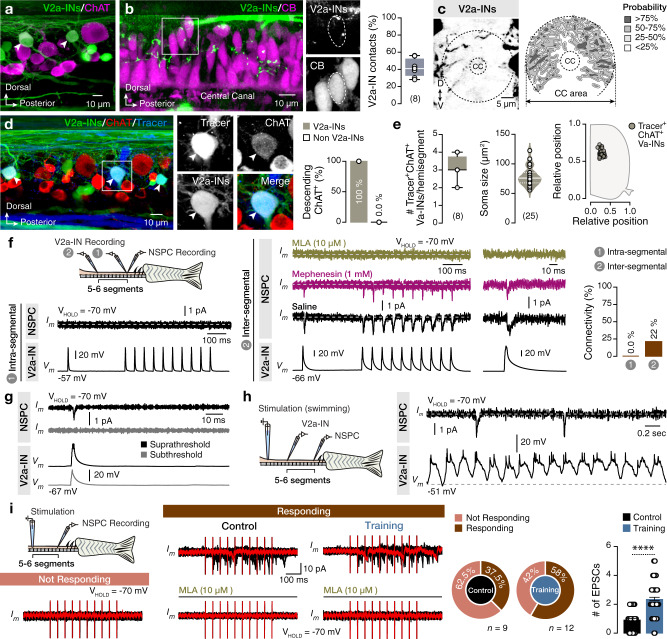
Fig. 4 Spinal locomotor V2a-INs contribute cholinergic inputs to NSPCs.

a Large and dorsally located spinal cord V2a-INs (Chx10:GFP+, green) are cholinergic (ChAT+, magenta). Arrowheads indicate double-labeled neurons (Chx10:GFP+ChAT+). b A sample stack from the central canal area showing the presence of V2a-IN (Chx10:GFP+) axonal collaterals (green) close to CB+ NSPCs (magenta) with analysis of the proportion of the CB+ NSPCs that are in close proximity with the V2a-IN (GFP+) processes. c Quantification of the probability of the V2a-IN axonal collaterals in the central canal region (n = 15 zebrafish). d Representative whole-mount confocal image showing that all (25 out of 25; 100% from 8 zebrafish) long descending (dextran tracer, blue; >10 segments) cholinergic (ChAT+, red) neurons are V2a-INs (GFP+, green). Arrowheads indicate triple-labeled neurons. e Quantification and analysis of the number, size and location of long descending cholinergic V2a-INs in the adult zebrafish spinal hemisegments. f Sample average (~25 sweeps) traces from dual electrophysiological recordings between a premotor V2a-IN and NSPCs, located in the same segment (intra-segmental, 1) or 5–6 segments rostrally (inter-segmental, 2). Cholinergic connections were observed in the inter-segmental pairs (22%, 11 out of 50 pairs) but not in the intra-segmental pairs (0%, 0 out of 15). g Postsynaptic responses in the recorder NSPCs generated from suprathreshold (black, with action potential) and not from subthreshold (gray, without an action potential) short pulse depolarization of the V2a-IN. h Ex vivo setup of the brain-spinal cord preparation allows simultaneous recordings of a NSPC and ipsilateral descending V2a-INs during fictive locomotion. Sample trace of a connected pair that, while the V2a-IN discharges during fictive swimming, the NSPC receives occasional input. i Illustration of recordings acquired during electrical stimulations. Ten pulses (20 Hz) of a rostral spinal cord segment were applied to depolarize V2a-INs connected to NSCs. Representative sample trace (in red) of superimposed sweeps (~20, in black) from not responding and responding NSPCs. Bath application of the selective nicotinic antagonist MLA (10 μM) abolished the recorded currents suggesting that they are cholinergic. Changes in the proportion of the recorded NSPCs that respond to electrical stimulation observed after training (n: number of recorded NSPCs). The average number of detected events per stimulation sweep from both sites was significantly higher in trained animals, suggesting adaptive changes in the innervation and the cholinergic release to the NSCs (P < 0.0001). The dashed gray line represents the baseline. CB, calbindin D-28K; CC, central canal; ChAT, choline acetyltransferase; EPSC, excitatory postsynaptic current; INs, interneurons; MLA, methyllycaconitine; NSPC, neural stem/progenitor cell. Data are presented as mean ± s.e.m., as violin plots and as box plots showing the median with 25/75 percentile (box and line) and minimum–maximum (whiskers). ****P < 0.0001. For detailed statistics, see Supplementary Table 1.

Acknowledgments
This image is the copyrighted work of the attributed author or publisher, and ZFIN has permission only to display this image to its users. Additional permissions should be obtained from the applicable author or publisher of the image. Full text @ Nat. Commun.