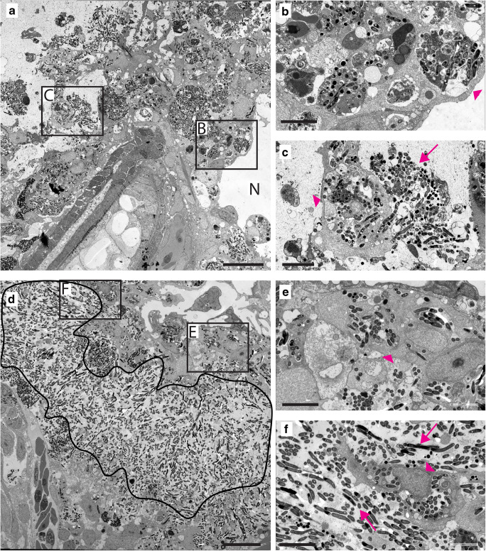
IMAGE

Fig. 4

ID
ZDB-IMAGE-210821-34
Source
Figures for Hosseini et al., 2021
Image
Figure Caption

Fig. 4

Granuloma structures in myd88-/- larvae consist mainly of extracellular Mm. a TEM image of a granuloma in representative myd88+/+ larvae, showing the necrotic centre (N) and aggregates of Mm in the immune cells. b,c Higher magnification of regions indicated in (a), showing infected cells (arrowheads) and extracellular Mm (arrows). d TEM image of a granuloma in representative myd88-/- larvae showing the area with extracellular Mm (black line). e,f Higher magnification of region indicated in (d), showing extracellular bacteria and infected cells (arrowheads). n = 3 per group, the scale bars in (a)–(d) (20 μm) and in (b)–(c) and (e)–(f) (10 μm)

Figure Data
Acknowledgments
This image is the copyrighted work of the attributed author or publisher, and ZFIN has permission only to display this image to its users. Additional permissions should be obtained from the applicable author or publisher of the image. Full text @ Virchows Arch