
Figure 3
- ID
- ZDB-IMAGE-210801-98
- Publication
- Quadri et al., 2021 - Phosphorylation of H3-Thr3 by Haspin Is Required for Primary Cilia Regulation
- All Figures
- Figures for Quadri et al., 2021
|
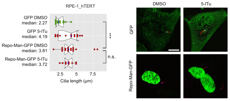
Figure 3 Loss of H3-Th3p causes an increase in cilia length. RPE-1_hTERT cells were transfected with a GFP- or Repo-Man-GFP- encoding plasmid and serum-starved for 24 h before being incubated, in serum-free media, with DMSO or 10 nM 5-iTU for a further 24 h. Cells were then fixed and processed for immunofluorescence against γ-tubulin (pink) and acetylated-tubulin (red). Representative images are shown (scale bar: 10 µm). Graph shows the median cilia length calculated as described in Material and Methods; boxes include 50% of the data points, notch represent confidence interval (median ± 1.58 IQR/sqrt(n)). t-test was applied as a statistical measurement, n.s.; not significant, *** p < 0.005.