
Figure 1
- ID
- ZDB-IMAGE-210801-8
- Genes
- Publication
- Li et al., 2021 - Disruption of MAP7D1 Gene Function Increases the Risk of Doxorubicin-Induced Cardiomyopathy and Heart Failure
- All Figures
- Figures for Li et al., 2021
|
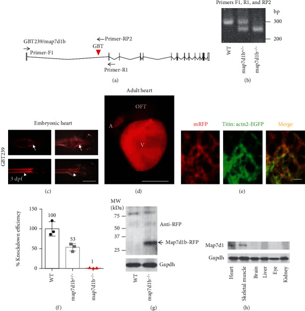
Figure 1 A transposon insertion in the GBT239 mutant disrupted the map7d1b gene that is predominantly expressed in the cardiac and skeletal muscle. (a) Insertional position of a gene-break transposon (GBT) element into the first intron of the map7d1b gene in the GBT239 mutant. (b) Representative DNA gel images of PCR genotyping for identifying GBT239 heterozygous (map7d1b+/-) and GBT239 homozygous (map7d1b-/-) mutant alleles. (c) Imaging the GBT239 mutant at 3 days postfertilization (dpf) reported the cardiac (arrows) and skeletal muscle (arrowheads) specific expression of the tagged Map7d1b protein. Scale bar: 0.5 mm. (d, e) Imaging the GBT239 adult heart indicated ventricle enriched expression of the tagged Map7d1b protein (d), which largely overlapped with the EGFP signal in the sarcomere reporter line Tg(titin:actn2-EGFP) (e). V: ventricle; A: atrium; OFT: outflow tract. Scale bar in (d): 1 mm. Scale bar in (e): 20 μm. (f) Quantitative RT-PCR analysis of the native map7d1b transcript disruption in the GBT239/map7d1b mutant. (g) Western blotting analysis of the Map7d1b-RFP fusion protein in the GBT239/map7d1b mutant. Arrow indicates the predicted size of Map7d1b-RFP fusion protein. (h) Western blotting analysis of the Map71b protein in different mouse tissues.