IMAGE

Fig 7

ID
ZDB-IMAGE-210801-52
Antibodies
Source
Figures for Vong et al., 2021
Image
Figure Caption

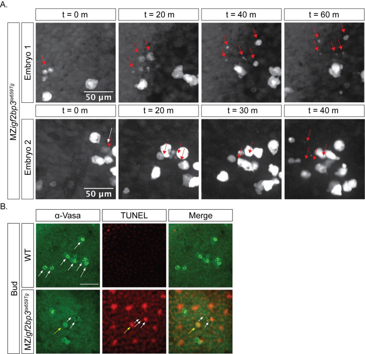
Fig 7 Loss of PGCs in MZ<italic toggle='yes'>igf2bp3</italic><sup><italic toggle='yes'>la659Tg</italic></sup> mutant embryos might arise from cell death.

A. Some PGCs are lost during migration in maternal igf2bp3la659Tg embryos. At bud stage, a subset of PGCs labelled with a GFP-nos1 live reporter display fragmentation during migration. B. Immunofluorescence with an α-Vasa antibody and TUNEL staining showed that compared to wild type embryos, in MZigf2bp3la659Tg embryos a subset of PGCs undergo cell death and fragmentation at the bud stage. Yellow arrow indicates co-localisation of Vasa and TUNEL labelling in a PGC, white arrows indicate Vasa+ fragments; Scale bar, 50 μm.

Figure Data
Acknowledgments
This image is the copyrighted work of the attributed author or publisher, and ZFIN has permission only to display this image to its users. Additional permissions should be obtained from the applicable author or publisher of the image. Full text @ PLoS Genet.