IMAGE

FIGURE 3

ID
ZDB-IMAGE-210729-26
Source
Figures for Tang et al., 2021
Image
Figure Caption

FIGURE 3

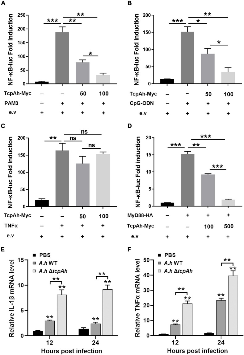
Examination of the inhibitory role of TcpAh in MyD88 signaling pathway. (A–C) Activation of the NF-κB-binding promoters detected in zebrafish embryos microinjected with NF-κB luciferase reporter (NF-κB-Luc; 100 pg/embryo), renilla luciferase reporter (10 pg/embryo), and increasing amounts (0, 50, and 100 pg/embryo) of TcpAh expression vectors with stimulation of (A) Pam3CSK4 (200 pg/embryo), (B) CpG-ODN (400 pg/embryo), and (C) TNFα (10 pg/embryo) for 12 h. Data are the average luciferase activity ± SD (*p < 0.05; **p < 0.01; ***p < 0.001; ns, not significant). (D) Activation of the NF-κB-binding promoter detected in HEK293T cells transfected with NF-κB luciferase reporter (NF-κB-Luc; 150 ng/mL), renilla luciferase reporter (15 ng/mL), MyD88 expression vector (50 ng/mL), and increasing amounts (0, 100, and 500 ng/mL) of TcpAh expression vectors. Data are the average luciferase activity ± SD (**p < 0.01; ***p < 0.001). (E,F) Real-time PCR analysis for the expression of zebrafish IL-1β (E) and TNFα (F) in leukocytes, which were sorted from peripheral blood, spleen, and kidney tissues at indicated time after i.p. stimulation with PBS, wild-type A. hydrophila and ΔtcpAh mutant. Data are representative of three independent experiments as mean ± SD (**p < 0.01). Standard loading was indicated by β-actin expression.

Acknowledgments
This image is the copyrighted work of the attributed author or publisher, and ZFIN has permission only to display this image to its users. Additional permissions should be obtained from the applicable author or publisher of the image. Full text @ Front Microbiol