IMAGE

FIGURE 2

ID
ZDB-IMAGE-210729-25
Source
Figures for Tang et al., 2021
Image
Figure Caption

FIGURE 2

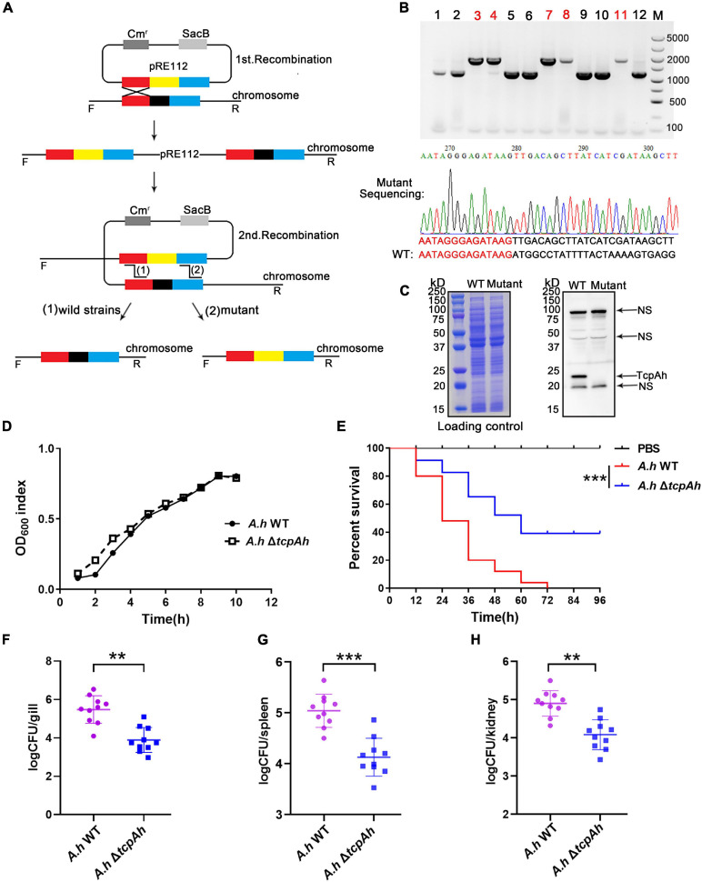
Examination of the requirement of TcpAh for A. hydrophila infection. (A) Strategy for the deletion of the tcpAh gene in A. hydrophila JBN2301 strain by homologous recombination. The black block indicates the tcpAh gene; red and blue blocks indicate the left and right homologous sequences of the tcpAh gene, respectively; and the yellow block indicates the Tcr gene. (B) PCR and sequencing identification of the mutant strain with tcpAh gene deletion (ΔtcpAh). The lanes with red number indicate the mutant strain; partial sequencing result is showed. (C) Western blot analysis of TcpAh protein in wild-type and mutant A. hydrophila JBN2301 strains in liquid cultures and NS means non-specific band. The total proteins of wild-type and ΔtcpAh A. hydrophila JBN2301 strains were separated by SDS-PAGE and stained with Coomassie brilliant blue R250 as a loading control. The TcpAh protein was detected by using a polyclonal rabbit anti-TcpAh antibody with an expected molecular weight of 22 kDa. (D) Growth curve of wild-type and ΔtcpAh A. hydrophila JBN2301 strains. (E) Zebrafish survival curve. Zebrafish were infected with wild-type A. hydrophila JBN2301 and ΔtcpAh mutant. Statistical differences between wild-type versus ΔtcpAh A. hydrophila infected groups were analyzed by log-rank test. n = 25. ***p < 0.001. Group of fish injected with mock PBS was used as a negative control. (F–H) Examination of bacterial load in the (F) gill, (G) spleen, and (H) kidney tissues of zebrafish infected with wild-type and ΔtcpAh mutant A. hydrophila JBN2301 strains. Non-parametric two-tailed Mann–Whitney test was carried out with (F)∗∗p < 0.01, (G)***p < 0.001, and (H) **p < 0.01.

Acknowledgments
This image is the copyrighted work of the attributed author or publisher, and ZFIN has permission only to display this image to its users. Additional permissions should be obtained from the applicable author or publisher of the image. Full text @ Front Microbiol