
|
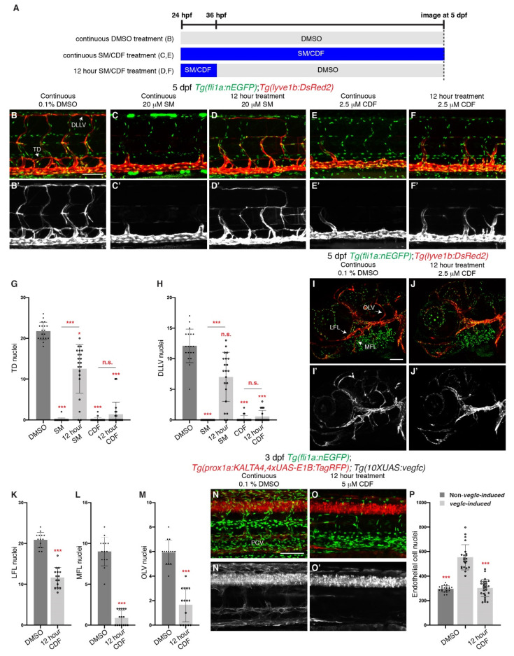
Figure 5 Brief treatment of 3,4-Difluorobenzocurcumin is sufficient to completely inhibit trunk and facial lymphatic development. (A) Schematic representation of the treatment schedule for larvae in images (B–F) and images (I,J). (B–F’) Lateral confocal images of 5 dpf Tg(fli1a:nEGFP);Tg(-5.2lyve1b:DsRed2) larvae either continuously treated with 0.1% DMSO (B,B’), 20 μM sunitinib malate (SM, C,C’) or 2.5 μM 3,4-Difluorobenzocurcumin (CDF, E,E’), or treated for 12 h with 20 μM SM (D,D’) or 2.5 μM CDF (F,F’). 12 h treatment of CDF inhibits trunk lymphatic development. Images (B’–F’) represent the Tg(-5.2lyve1b:DsRed2) expression of images (B–F). (G,H) Quantification of thoracic duct (TD, G) or dorsal longitudinal lymphatic vessel (DLLV, H) nuclei across 9 somites in 5 dpf Tg(fli1a:nEGFP);Tg(-5.2lyve1b:DsRed2) larvae treated with either 0.1% DMSO (n = 19 larvae), 20 μM SM (n = 24 larvae) or 2.5 μM CDF (n = 21 larvae), or treated for 12 h with 20 μM SM (n = 21 larvae) or 2.5 μM CDF (n = 21 larvae). (I–J’) Lateral confocal images of Tg(fli1a:nEGFP);Tg(-5.2lyve1b:DsRed2) larvae treated with either 0.1% DMSO (I,I’) or with 2.5 μM CDF for 12 h, then with 0.1% DMSO up to 5 dpf (J,J’). 12 h treatment of CDF inhibits facial lymphatic development. Images (I’,J’) represent the Tg(-5.2lyve1b:DsRed2) expression of images (I,J). (K–M) Quantification of lateral facial lymphatic (LFL, K, n ≥ 14), medial facial lymphatic (MFL, L, n ≥ 14), or otolithic lymphatic vessel (OLV, M, n ≥ 14) nuclei in 5 dpf Tg(fli1a:nEGFP);Tg(-5.2lyve1b:DsRed2) larvae either continuously treated with 0.1% DMSO (n = 14 larvae) or treated for 12 h with 2.5 μM CDF (n = 15 larvae). Datasets for 0.1% DMSO-treated 5 dpf Tg(fli1a:nEGFP);Tg(-5.2lyve1b:DsRed2) larvae are taken from Figure 1P–R. (N–O’) Lateral confocal images of 3 dpf Tg(prox1a:KALTA4,4xUAS-E1B:TagRFP);Tg(10XUAS:vegfc);Tg(fli1a:nEGFP) larvae (vegfc-induced) treated with either 0.1% DMSO (N,N’) or with 5 μM CDF for 12 h, then with 0.1% DMSO up to 3 dpf (O,O’). Pathological vascular phenotypes in vegfc-induced embryos are rescued by 12 h treatment of CDF. Images (N’,O’) represent the Tg(prox1a:KALTA4,4xUAS-E1B:TagRFP) expression of images (N,O). To avoid the robust prox1a expression in muscle cells, (N’,O’) are maximum projection images of only the z stacks that contain the posterior cardinal vein. Image (N’) (21/22 embryos) shows an embryo with increased prox1a:KALTA4,4xUAS-E1B:TagRFP expression in venous endothelial cells. This pathological phenotype is rescued in image (O’) (24/27 embryos). (P) Quantification of fli1a:EGFP-positive ECs across 4.5 somites in either 3 dpf Non-vegfc-induced (n = 21 embryos) or 3 dpf vegfc-induced larvae treated with either 0.1% DMSO (n = 22 embryos), or for 12 h with 5 μM CDF (n = 27 embryos). Datasets for 0.1% DMSO-treated 3 dpf Non-vegfc induced and vegfc-induced larvae are taken from Figure 4H. PCV: posterior cardinal vein. Statistical test: Mann-Whitney test were conducted for graph (K–M). Kruskal-Wallis test were conducted for graphs (G,H,P). p ≤ 0.001 (***) and n.s. indicates not significant. Scale bars: 100 μm.