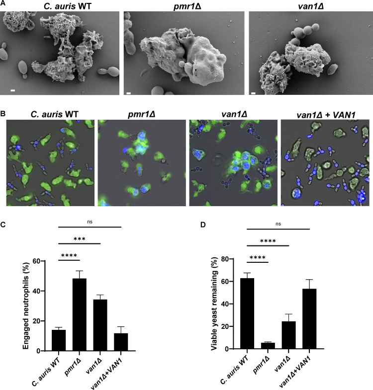
FIG 2 C. auris mannosylation pathway mutants are susceptible to neutrophil attack. (A) C. auris strains were incubated with human neutrophils for 1 h and were subsequently imaged via scanning electron microscopy. Images are 10,000× magnification, measurement bars represent 1 μm. (B and C) Human neutrophils were labeled with calcein-AM (green) and cocultured with individual C. auris strains labeled with calcofluor white (blue) for 1 h and imaged via fluorescence microscopy (B). The numbers of neutrophils engulfing fungal cells were counted and the percentages of total engaged neutrophils were calculated (C); n ≥ 3, mean with standard error of the mean (SEM) shown. (D) Individual C. auris strains were cultured with human neutrophils for 4 h and viable burden was estimated by PrestoBlue metabolic activity following neutrophil lysis; n = 3, mean with standard deviation shown. *, P < 0.05; **, P < 0.01; ***, P < 0.001; ****, P < 0.0001; ns, not significant by one-way ANOVA with Holm-Sidak multiple comparisons to C. auris WT.
Image
Figure Caption
Acknowledgments
This image is the copyrighted work of the attributed author or publisher, and
ZFIN has permission only to display this image to its users.
Additional permissions should be obtained from the applicable author or publisher of the image.
Full text @ mSphere

