
Figure 5
- ID
- ZDB-IMAGE-210708-29
- Antibodies
- Publication
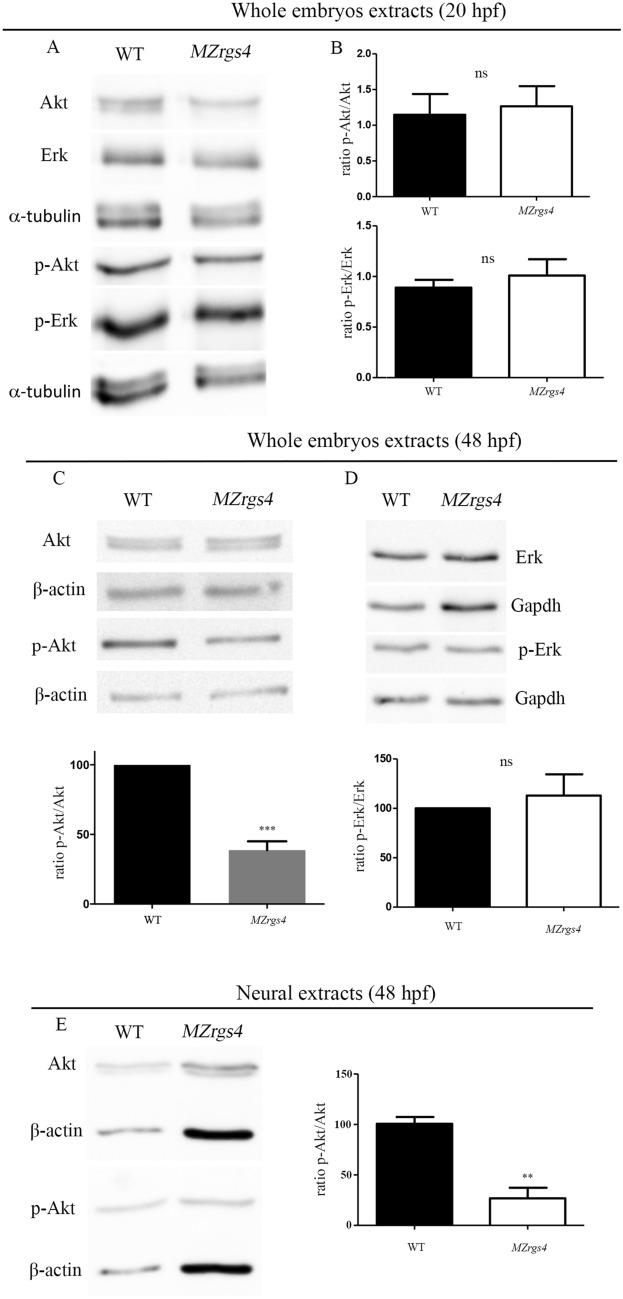
- Mikdache et al., 2021 - Rgs4 is a regulator of mTOR activity required for motoneuron axon outgrowth and neuronal development in zebrafish
- All Figures
- Figures for Mikdache et al., 2021
|
Figure 5
Loss of