
|
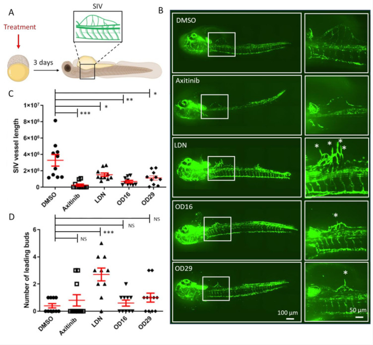
Figure 6 OD16 and OD29 inhibit subintestinal vessel (SIV) formation in transgenic Casper zebrafish line Tg (fli:EGFP). (A) Schematic representation of the experimental procedure. Freshly collected 2.5 hpf embryos were challenged with testing compounds in egg water. The SIV vascularization was analyzed after 3 days of treatment. (B) Effects of OD16, OD29, Axitinib, and LDN-193189 at 0.5 µM, or vehicle control (DMSO) on zebrafish SIV development. Compounds were added to egg water for 3 days before observing the SIV vascularization. Representative images of overview (left panel) and high magnification (white rectangle areas from left) are shown. Asterisks represent sprouting vessels. Scale bars represent 100 and 50 µm. (C) Quantification of the SIV vessel length of the zebrafish embryos in each group from the experiment shown in (B). * p < 0.05, ** p < 0.005, *** p < 0.001. (D) Quantification of the number of leading buds (indicated as asterisks in panel B). *** p < 0.001. NS, not significant.