IMAGE

Figure 3

ID
ZDB-IMAGE-210628-48
Source
Figures for Ishii et al., 2021
Image
Figure Caption

Figure 3

Opacification of paraffin blocks using white agarose gel. Specimens are pre-embedded in agarose gel containing white watercolor, then dehydrated and embedded in paraffin. (
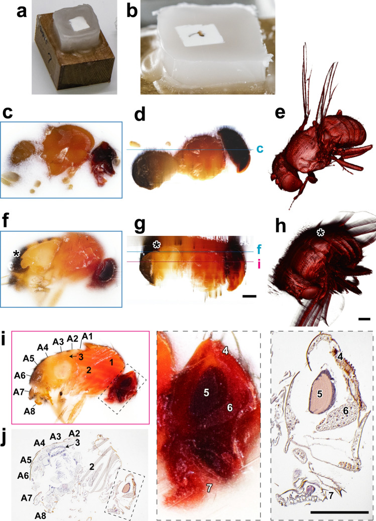
a) A fly was fixed and pre-embedded in white agarose, then dehydrated and embedded in paraffin. (b) In the block face, white agarose gives opaque area around the specimen, while pure paraffin is seen as transparent area around the lump of white agarose. (ce) A fly pre-embedded in white agarose generated 276 block-face images with 4 µm-interval. A block-face image (c), a reconstructed (d), and a volume-rendered image (e) are shown. (fh) Without white agarose, structures underneath the block face are visible through the paraffin in the block-face image (* in f), and a reconstructed plane (* in g). These structures are represented in the 3D volume-rendered image as a shadow (* in h). Images in (g) and (h) were reconstructed from 351 block-face images with 4 µm-interval. (i, j) Correlation between a block-face image (i) and microscopic image of a section (j) were performed using the specimen shown in (fh). Boxed areas in (i) and (j) are magnified in the right panels. A1–A8: Abdominal segments, 1: Dorso-longitudinal muscles, 2: Dorso-ventral muscles, 3: Midgut, 4: Compound eyes, 5: Brain, 6: Head fat body, 7: Labellum. Scale bars: 200 µm.

Acknowledgments
This image is the copyrighted work of the attributed author or publisher, and ZFIN has permission only to display this image to its users. Additional permissions should be obtained from the applicable author or publisher of the image. Full text @ Sci. Rep.