
|
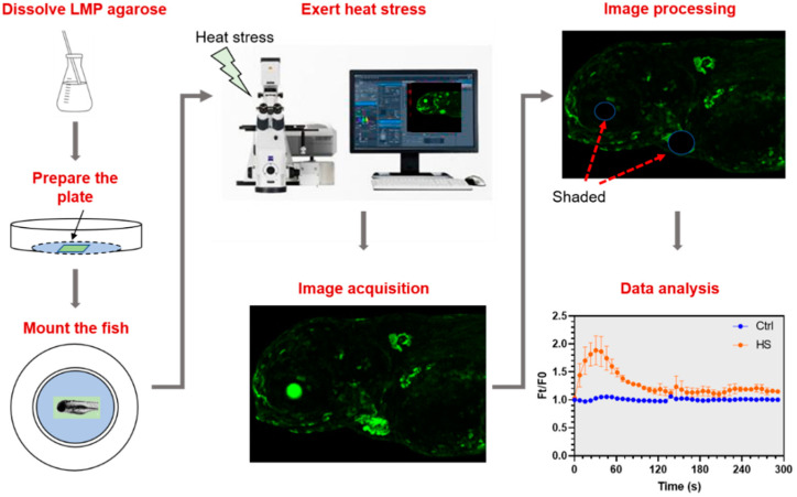
Figure 3 Workflow of the protocol to analyze in vivo activities of Ca2+ signaling triggered by heat stress. Low melting-point (LMP) agarose was melted and poured into a confocal dish. After solidification of the LMP agarose, a tiny pit was made in the middle of the agarose plane. A larva anesthetized with MS-222 was put into the pit. The dish was placed onto the stage of a confocal microscope and orientation of the larvae was adjusted. After that, Z-stack time-lapse imaging was initiated immediately upon addition of hot water into the dish. Taking into account strong basal fluorescence of the eyes and the heart, these organs were shaded before measuring fluorescence intensity. Finally, the time-lapse fluorescence intensity data were plotted to illustrate the dynamics of Ca2+ signaling elicited by heat stress.